. 2010 Mar;58(3):640-9.
doi: 10.1016/j.neuropharm.2009.11.013. Epub 2009 Dec 4.

Pharmacological activity of C10-substituted analogs of the high-affinity kainate receptor agonist dysiherbaine

Affiliations

Pharmacological activity of C10-substituted analogs of the high-affinity kainate receptor agonist dysiherbaine

L Leanne Lash-Van Wyhe et al. Neuropharmacology. 2010 Mar.

Abstract

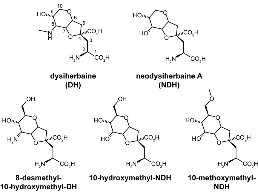
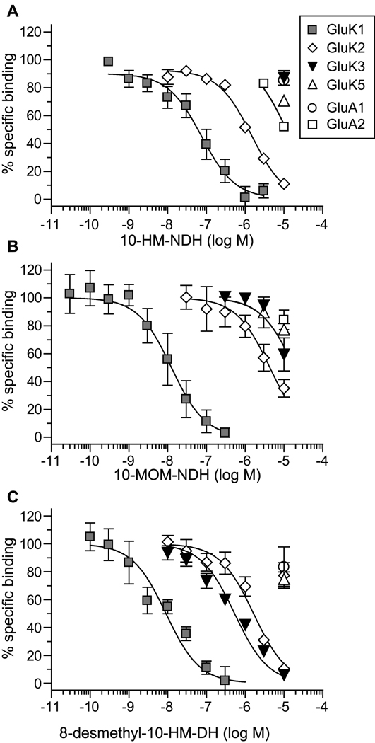
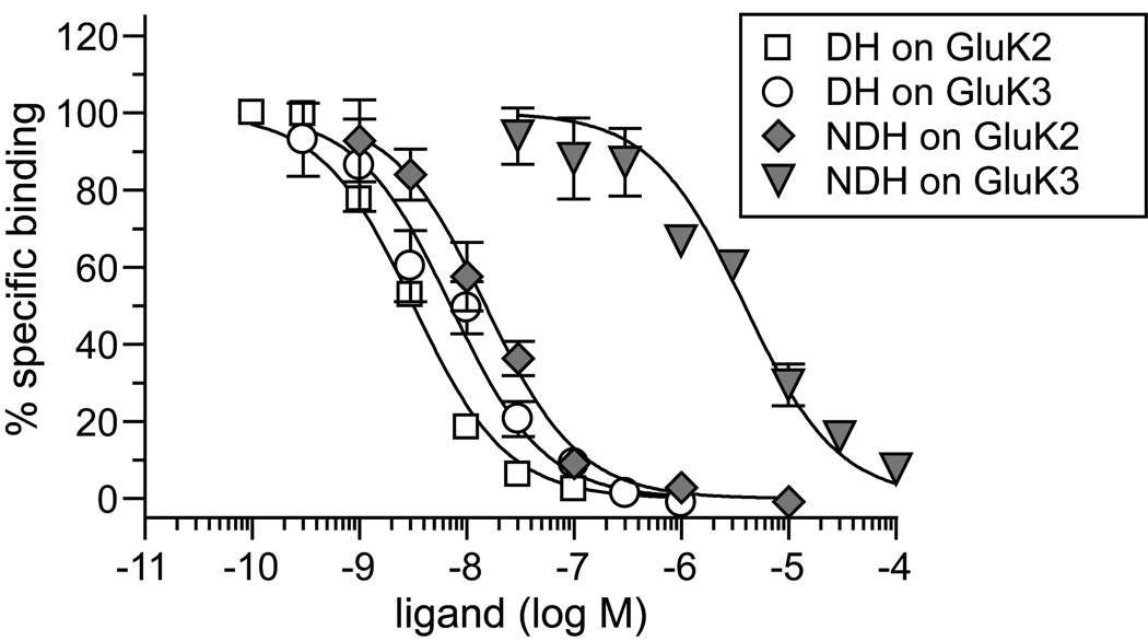
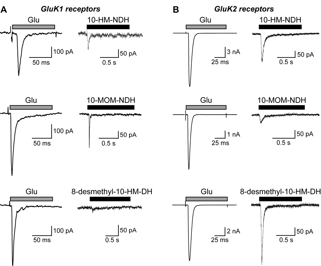
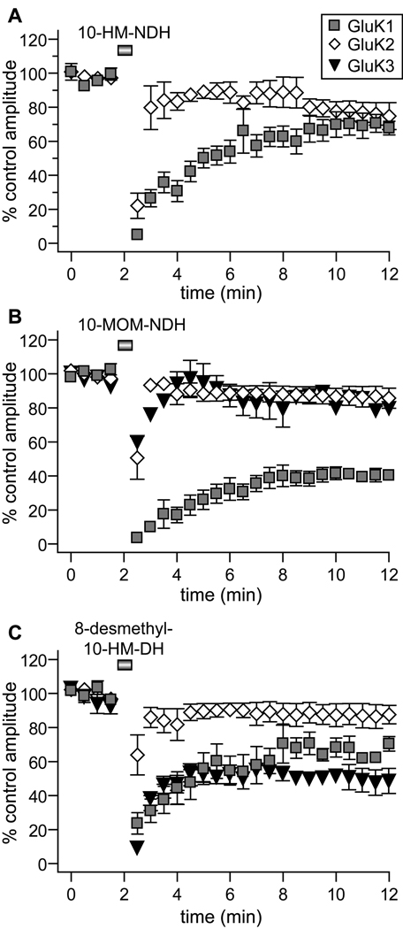
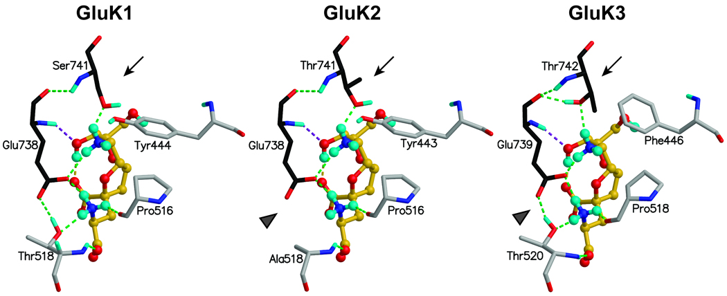
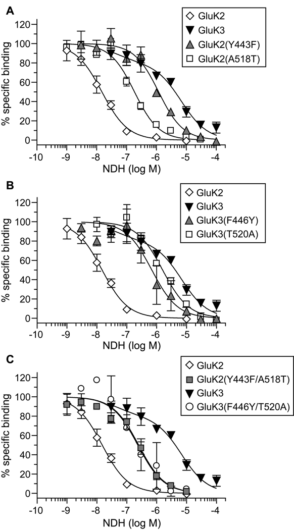
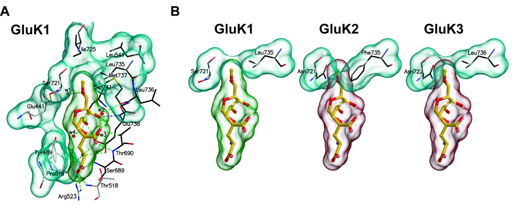
Kainate receptor antagonists have potential as therapeutic agents in a number of neuropathologies. Synthetic modification of the convulsant marine toxin neodysiherbaine A (NDH) previously yielded molecules with a diverse set of pharmacological actions on kainate receptors. Here we characterize three new synthetic analogs of NDH that contain substituents at the C10 position in the pyran ring of the marine toxin. The analogs exhibited high-affinity binding to the GluK1 (GluR5) subunit and lower affinity binding to GluK2 (GluR6) and GluK3 (GluR7) subunits in radioligand displacement assays with recombinant kainate and AMPA receptors. As well, the natural toxin NDH exhibited approximately 100-fold selectivity for GluK2 over GluK3 subunits, which was attributable to the C8 hydroxyl group in NDH. We used molecular dynamic simulations to determine the specific interactions between NDH and residues within the ligand-binding domains of these two kainate receptor subunits that contribute to the divergent apparent affinities for the compound. These data demonstrate that interactions with the GluK1 subunit are preserved in analogs with substitutions at C10 in NDH and further reveal the determinants of selectivity and pharmacological activity of molecules acting on kainate receptor subunits, which could aid in design of additional compounds that target these receptors.


Figures

Figure 1
Figure 1
Two dimensional structures of the synthetic C10 analogs of DH. The parent marine toxins, dysiherbaine (DH) and neodysiherbaine (NDH) are shown in the top row of structures. Numbering of the carbons is shown for the DH structure.
Figure 2
Figure 2
C10 analogs of DH bind with highest affinity to GluK1 subunits in radioligand displacement assays. Displacement of [3H]kainate or [3H]AMPA from kainate and AMPA receptor subunits, respectively, for (A) 10-HM-NDH, (B) 10-MOM-NDH, and (C) 8-desmethyl-10-HM-DH. Symbols representing data from GluK1–3, GluK5, and GluA1–2 subunits are as shown in the boxed key in A. Data was fit with a one-site competition curve with fixed minima (0%) and maxima (100%) (n = 3–5 for each concentration of analog on each receptor subunit). Ki values derived from this data are given in Table 1.
Figure 3
Figure 3
NDH, but not DH, shows ∼100-fold selectivity for GluK2 over GluK3 subunits. Displacement of [3H]kainate from GluK2 and GluK3 subunits by DH and NDH. The competition curve for DH on GluK2 is from previously published data and is shown only for the sake of comparison (Sakai et al., 2001b). Data was fit with a one-site competition curve with fixed minima (0%) and maxima (100%) (n = 3–5 for each concentration of analog on each receptor subunit). Ki values derived from this data are given in Table 1.
Figure 4
Figure 4
The C10 analogs of DH are kainate receptor agonists. (A). Representative GluK1 receptor whole-cell currents evoked by application of glutamate (10 mM, 100 ms) or the C10 analogs (30 µM, 1 sec). (B). Representative currents from GluK2 receptors with the same set of agonists. Cells were held at −70 mV, lifted from the coverslip into a laminar solution stream, and agonists were fast-applied with a piezoceramic translation system. Traces are representative of at least 3 recordings under each condition.
Figure 5
Figure 5
Time course of recovery of glutamate-evoked currents after application of C10 analogs to kainate receptors. The data represent the normalized amplitude of glutamate-evoked currents (10 mM, 100 ms) relative to the control two-minute period before receptors were exposed to the C10 analogs. (A) 10-HM-NDH, (B) 10-MOM-NDH, and (C) 8-desmethyl-10-HM-DH were applied at 30 µM for 30 sec. Glutamate applications were then resumed at 20 sec intervals for the duration of the experiment. (n = 3–6 at each time point). In each case, GluK1 receptors recovered from analog-induced desensitization at the slowest rate; in the case of 10-MOM-NDH, amplitudes plateaued at a level lower than that of the control. GluK2 receptors recovered quickly in each case. 10-HM-NDH was not analyzed on GluK3 receptors in (A) because this analog showed no affinity for the GluK3 receptor (see Table 1).
Figure 6
Figure 6
Binding of 8-desmethyl-10-HM-DH to GluK1-, GluK2-, and GluK3-LBCs derived from MD simulations. Carbon atoms of the amino acid residues in lobe D1 are colored with grey, and from lobe D2 with black. The ligand is shown with an yellow carbon atoms. The hydrogen-bonding interactions are shown with dotted lines. Polar hydrogens (cyan) are shown, while those attached to carbon atoms are left out for clarity. The arrow marks the site of Ser/Thr741-Tyr/Phe444 shift in conformation. The grey arrowhead denotes the position of the differential Ala/Thr518-Glu738 interaction. Numbering in each ligand binding domain is according to position in the full-length subunit.
Figure 7
Figure 7
Analysis of the effect of reciprocal mutations of Tyr443/Phe446 and Ala518/Thr520 on binding to NDH. A. Single site mutations in GluK2 of Tyr443 (Y443) to a phenylalanine and Ala518 (A518) to a threonine greatly reduced binding affinity, as evidenced by the substantial shifts to the right in the displacement curves. GluK2 and GluK3 displacement curves are shown for the sake of references in this and the following panels. B. Single mutations of Phe446 (F446) and Thr520 (T520) in GluK3 increased affinity for NDH only modestly. C. Double reciprocal mutation of residues 443/446 and 518/520 in GluK2 and GluK3 produced receptors with an identical displacement profile intermediate to that of the parent wildtype receptors. Ki values derived from this data are given in the text.
Figure 8
Figure 8
Binding mode of 10-MOM-NDH to kainate receptors derived from MD simulations with the GluK1 LBC. A. The detailed binding configuration for GluK1, demonstrating the compatibility of the C10 substitution for the hydrophobic niche in the binding pocket. B. The predicted effect of sequence differences in GluK1–3 at positions 721 and 735 in shaping the binding pocket and complementarity for C10 functional groups. Carbon atoms of the amino acid residues in D1 are colored with grey, and from D2 with black. The hydrogen bonding interactions are shown with dotted lines (see legend for Fig. 6 for additional information).

Similar articles

Cited by

References

    1. Alt A, Weiss B, Ogden AM, Li X, Gleason SD, Calligaro DO, Bleakman D, Witkin JM. In vitro and in vivo studies in rats with LY293558 suggest AMPA/kainate receptor blockade as a novel potential mechanism for the therapeutic treatment of anxiety disorders. Psychopharmacology (Berl) 2006;185:240–247. - PubMed
    1. Alt A, Weiss B, Ornstein PL, Gleason SD, Bleakman D, Stratford RE, Jr, Witkin JM. Anxiolytic-like effects through a GLU(K5) kainate receptor mechanism. Neuropharmacology. 2007;52:1482–1487. - PubMed
    1. Bayly C, Cieplak P, Cornell W, Kollman P. A well-behaved electrostatic potential based method using charge restraints for deriving atomic charges: the RESP model. J. Phys. Chem. 1993;97:10269–10280.
    1. Ben-Ari Y. Limbic seizure and brain damage produced by kainic acid: mechanisms and relevance to human temporal lobe epilepsy. Neuroscience. 1985;14:375–403. - PubMed
    1. Cieplak P, Cornell W, Bayly C, Kollman P. Application of the multimolecule and multiconformational RESP methodology to biopolymers-charge derivation for DNA, RNA, and proteins. J. Comput. Chem. 1995;16:1357–1377.

Publication types

MeSH terms