

UOK 262 cell line, fumarate hydratase deficient (FH-/FH-) hereditary leiomyomatosis renal cell carcinoma: in vitro and in vivo model of an aberrant energy metabolic pathway in human cancer
- PMID: 19963135
- PMCID: PMC2827193
- DOI: 10.1016/j.cancergencyto.2009.08.018
UOK 262 cell line, fumarate hydratase deficient (FH-/FH-) hereditary leiomyomatosis renal cell carcinoma: in vitro and in vivo model of an aberrant energy metabolic pathway in human cancer
Abstract
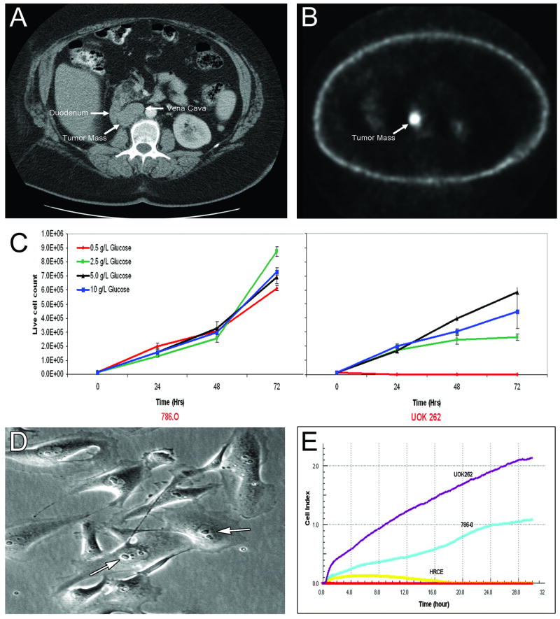
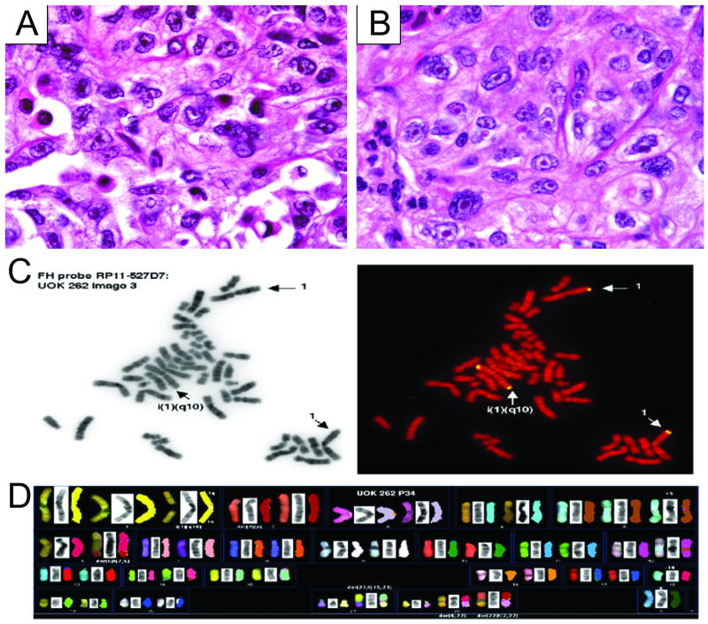
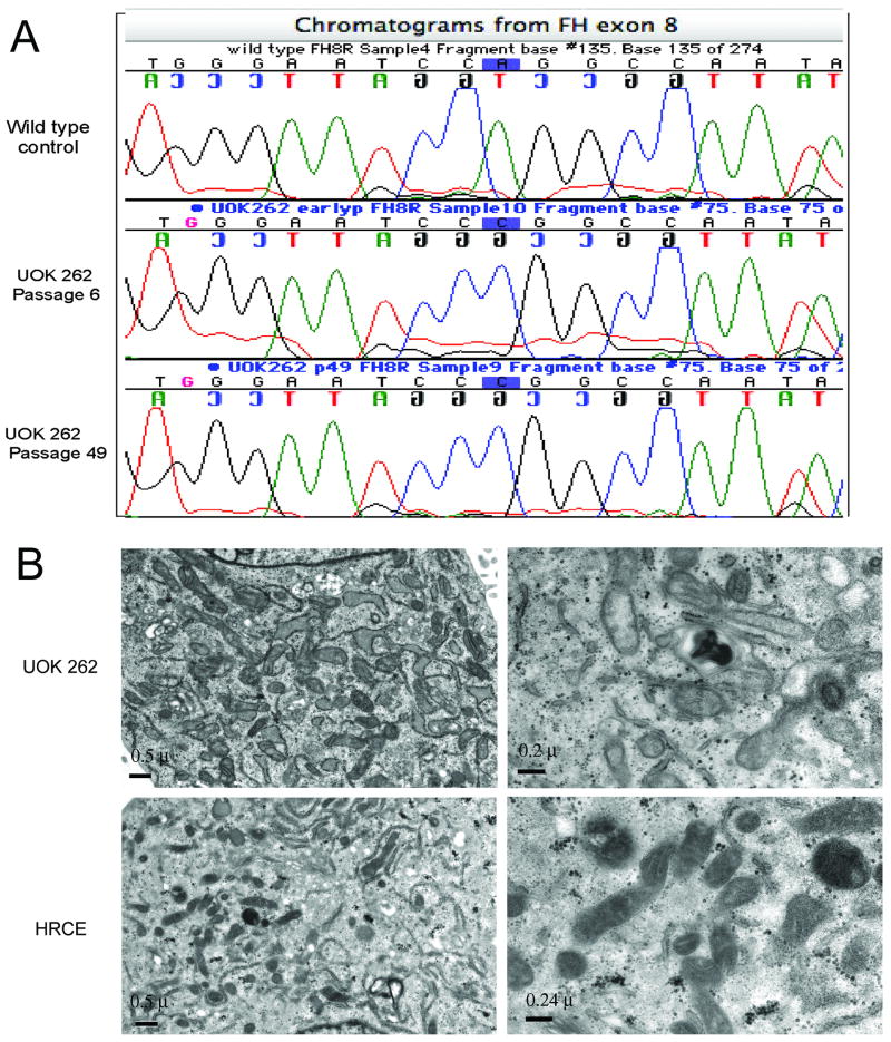
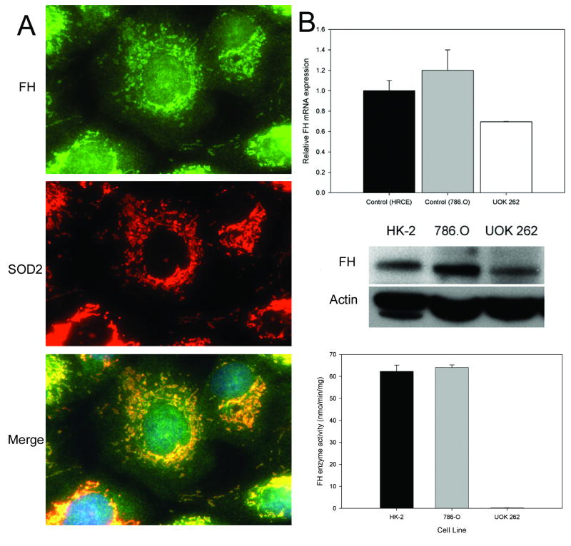
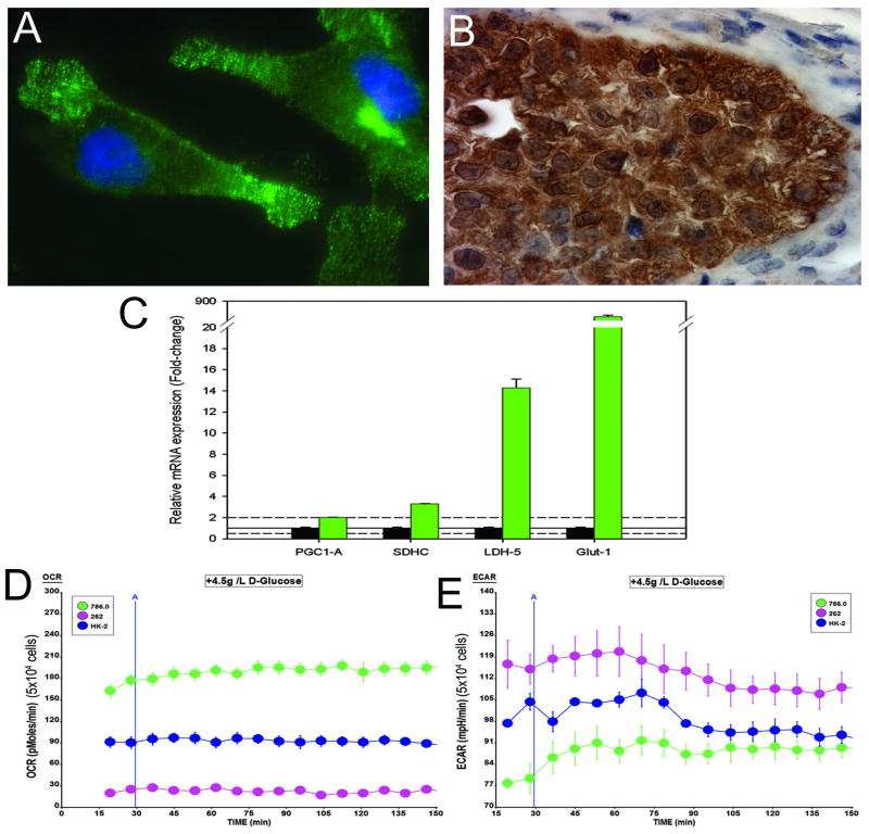
Energy deregulation and abnormalities of tumor cell metabolism are critical issues in understanding cancer. Hereditary leiomyomatosis renal cell carcinoma (HLRCC) is an aggressive form of RCC characterized by germline mutation of the Krebs cycle enzyme fumarate hydratase (FH), and one known to be highly metastatic and unusually lethal. There is considerable utility in establishing preclinical cell and xenograft models for study of disorders of energy metabolism, as well as in development of new therapeutic approaches targeting of tricarboxylic acid (TCA) cycle enzyme-deficient human cancers. Here we describe a new immortalized cell line, UOK 262, derived from a patient having aggressive HLRCC-associated recurring kidney cancer. We investigated gene expression, chromosome profiles, efflux bioenergetic analysis, mitochondrial ultrastructure, FH catabolic activity, invasiveness, and optimal glucose requirements for in vitro growth. UOK 262 cells have an isochromosome 1q recurring chromosome abnormality, i(1)(q10), and exhibit compromised oxidative phosphorylation and in vitro dependence on anaerobic glycolysis consistent with the clinical manifestation of HLRCC. The cells also display glucose-dependent growth, an elevated rate of lactate efflux, and overexpression of the glucose transporter GLUT1 and of lactate dehydrogenase A (LDHA). Mutant FH protein was present primarily in edematous mitochondria, but with catalytic activity nearly undetectable. UOK 262 xenografts retain the characteristics of HLRCC histopathology. Our findings indicate that the severe compromise of oxidative phosphorylation and rapid glycolytic flux in UOK 262 are an essential feature of this TCA cycle enzyme-deficient form of kidney cancer. This tumor model is the embodiment of the Warburg effect. UOK 262 provides a unique in vitro and in vivo preclinical model for studying the bioenergetics of the Warburg effect in human cancer.
Conflict of interest statement
Figures
References
-
- Fernandez-Pugnaire MA, gado-Florencio V. Familial multiple cutaneous leiomyomas. Dermatology. 1995;191:295–298. - PubMed
-
- Zbar B, Glenn G, Lubensky I, Choyke P, Walther MM, Magnusson G, Bergerheim US, Pettersson S, Amin M, Hurley K. Hereditary papillary renal cell carcinoma: clinical studies in 10 families. J Urol. 1995;153:907–912. - PubMed
-
- Tomlinson IP, Alam NA, Rowan AJ, Barclay E, Jaeger EE, Kelsell D, Leigh I, Gorman P, Lamlum H, Rahman S, Roylance RR, Olpin S, Bevan S, Barker K, Hearle N, Houlston RS, Kiuru M, Lehtonen R, Karhu A, Vilkki S, Laiho P, Eklund C, Vierimaa O, Aittomaki K, Hietala M, Sistonen P, Paetau A, Salovaara R, Herva R, Launonen V, Aaltonen LA. Germline mutations in FH predispose to dominantly inherited uterine fibroids, skin leiomyomata and papillary renal cell cancer. Nat Genet. 2002;30:406–410. - PubMed
-
- Kiuru M, Lehtonen R, Arola J, Salovaara R, Jarvinen H, Aittomaki K, Sjoberg J, Visakorpi T, Knuutila S, Isola J, Delahunt B, Herva R, Launonen V, Karhu A, Aaltonen LA. Few FH mutations in sporadic counterparts of tumor types observed in hereditary leiomyomatosis and renal cell cancer families. Cancer Res. 2002;62:4554–4557. - PubMed
Publication types
MeSH terms
Substances
Grants and funding
LinkOut - more resources
Full Text Sources
Other Literature Sources
Medical
Research Materials
Miscellaneous
