

Review
doi: 10.1016/j.ceb.2009.11.001.
Epub 2009 Dec 5.
Microtubule-severing enzymes
Affiliations
- PMID: 19963362
- PMCID: PMC2822099
- DOI: 10.1016/j.ceb.2009.11.001
Item in Clipboard
Review
Microtubule-severing enzymes
Curr Opin Cell Biol.
2010 Feb.
Abstract
In 1993, an enzyme with an ATP-dependent microtubule-severing activity was purified from sea urchin eggs and named katanin, after the Japanese word for sword. Now we know that katanin, spastin, and fidgetin form a family of closely related microtubule-severing enzymes that is widely distributed in eukaryotes ranging from Tetrahymena and Chlamydomonas to humans. Here we review the diverse in vivo functions of these proteins and the recent significant advances in deciphering the biophysical mechanism of microtubule severing.
Copyright 2009 Elsevier Ltd. All rights reserved.
Figures
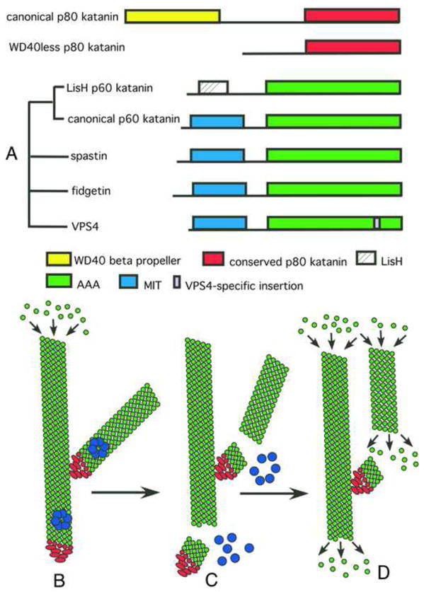
(A) Schematic representation of the domain architecture of microtubule-severing enzymes. Katanin purified from sea urchin eggs is a heterodimer with a regulatory, non-AAA subunit, p80 katanin, and a AAA catalytic subunit, p60 katanin. The phylogenetic relationship of proteins related to p60 katanin is indicated on the left. Based on their representation in sequenced genomes, we speculate that the LisH family of p60 katanins diverged early in eukaryotic evolution whereas the WD40less family of p80 katanins evolved independently in chordates and nematodes. (B–D) Schematic representation of branched nucleation and severing to generate parallel, treadmilling microtubule arrays in plant cells. (B) γ–tubulin ring complex (γTuRC), shown in red, binds to the wall of a pre-existing microtubule and nucleates polymerization of a new microtubule at a 40° angle. (C) Assembly of katanin rings, shown in blue, results in severing of these branched structures. (D) Severing by katanin frees microtubule minus ends to allow depolymerization of minus ends and allows branched arrays to re-arrange into parallel arrays.
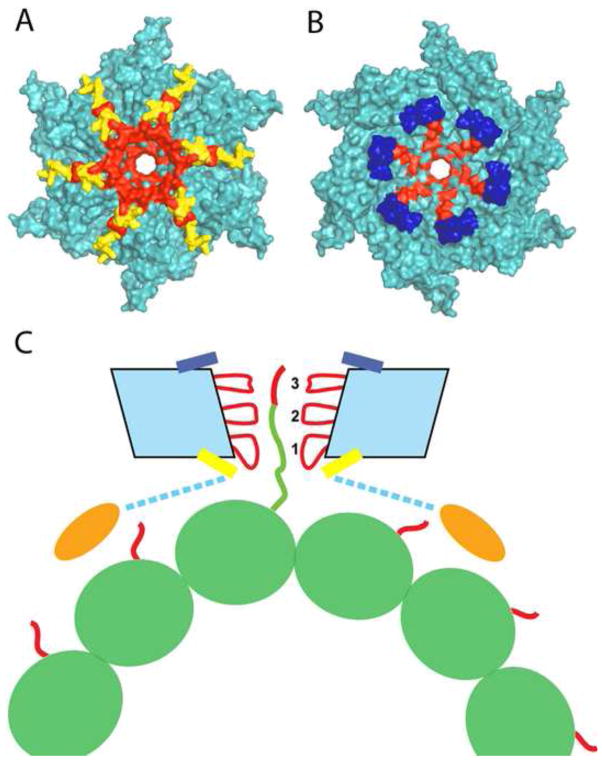
(A, B) Molecular surface of the spastin hexamer (after [39]). The N- and C-terminal helices that surround the entry and exit of the pore are shown in yellow and blue, respectively. Residues that line the entrance and the interior of the pore and are important for microtubule severing are shown in red. (C) Proposed mechanism for microtubule severing by spastin. The spastin AAA core is shown in cyan with Pore Loops 1,2 and 3 highlighted in red. The MIT domains are shown as gold ovals. The valency of the interaction of the MIT domains with the microtubule is unknown. Tubulin is shown in green, while the C-terminal tubulin tails are shown in red. Spastin uses its pore loops to exert “tugs” on the C-terminal tails and dislodge tubulin out of the microtubule lattice.
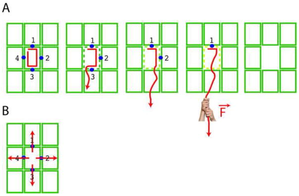
Schematic representation of the proposed mechanisms of microtubule severing for spastin and katanin (A) by sequential unfolding of the tubulin peptide chain, leading to gradual loss of the lattice contact points (blue dots) and complete removal of a tubulin subunit (green rectangle); (B) by prying apart the tubulin subunits in the microtubule lattice. The green rectangles represent the tubulin subunits within a small region of a microtubule.
References
-
- Hartman JJ, Mahr J, McNally K, Okawa K, Iwamatsu A, Thomas S, Cheesman S, Heuser J, Vale RD, McNally FJ. Katanin, a microtubule-severing protein, is a novel AAA ATPase that targets to the centrosome using a WD40-containing subunit. Cell. 1998;93:277–287. - PubMed
-
- Roll-Mecak A, Vale RD. The Drosophila homologue of the hereditary spastic paraplegia protein, spastin, severs and disassembles microtubules. Curr Biol. 2005;15:650–655. - PubMed
Publication types
MeSH terms
Substances
Grants and funding
LinkOut - more resources
Full Text Sources
Miscellaneous
