

Intraoperative demonstration of selective stimulation of the common human femoral nerve with a FINE
- PMID: 19963718
- PMCID: PMC3574574
- DOI: 10.1109/IEMBS.2009.5332757
Intraoperative demonstration of selective stimulation of the common human femoral nerve with a FINE
Abstract
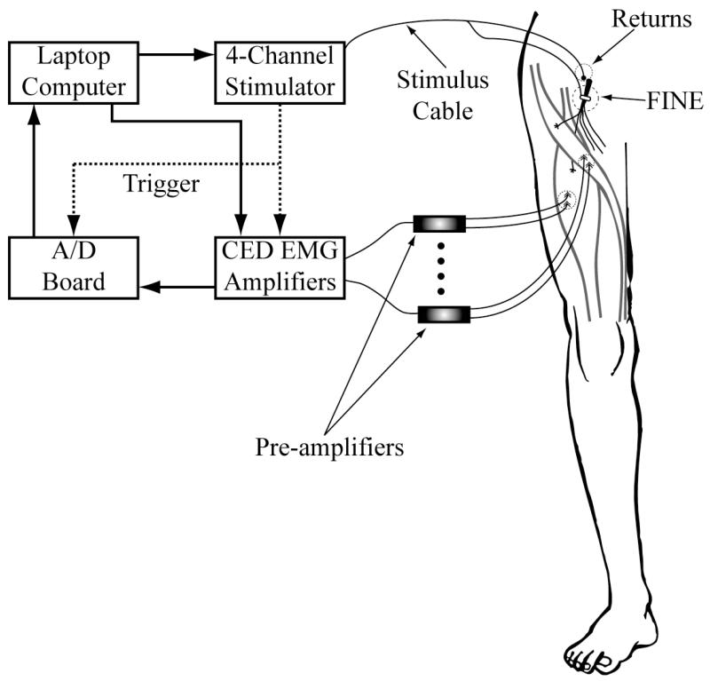
We have tested the hypothesis that the Flat Interface Nerve Electrode (FINE) can selectively stimulate each muscle innervated by the common femoral nerve of the human, near the inguinal ligament in a series of intraoperative trials. During routine vascular surgeries, an 8-contact FINE was placed around the common femoral nerve between the inguinal ligament and the first branching point. The efficacy of the FINE to selectively recruit muscles innervated by the femoral nerve was determined from electromyograms (EMGs) recorded in response to electrical stimulation. At least four of the six muscles innervated by the femoral nerve were selectively recruited in all subjects. Of these, at least one muscle was a hip flexor and two muscles were knee extensors. Results from the intraoperative experiments were used to estimate the potential for the electrode to restore knee extension and hip flexion through Functional Electrical Stimulation (FES). Normalized EMGs and biomechanical simulations were used to estimate joint moments and functional efficacy. Estimated knee extension moments exceed the threshold required for the sit-to-stand transition.
Figures


Similar articles
-
Selective stimulation of the human femoral nerve with a flat interface nerve electrode.J Neural Eng. 2010 Apr;7(2):26006. doi: 10.1088/1741-2560/7/2/026006. Epub 2010 Mar 8. J Neural Eng. 2010. PMID: 20208125 Free PMC article.
-
A model of selective activation of the femoral nerve with a flat interface nerve electrode for a lower extremity neuroprosthesis.IEEE Trans Neural Syst Rehabil Eng. 2008 Apr;16(2):195-204. doi: 10.1109/TNSRE.2008.918425. IEEE Trans Neural Syst Rehabil Eng. 2008. PMID: 18403289 Free PMC article.
-
Selective activation of the human tibial and common peroneal nerves with a flat interface nerve electrode.J Neural Eng. 2013 Oct;10(5):056006. doi: 10.1088/1741-2560/10/5/056006. Epub 2013 Aug 5. J Neural Eng. 2013. PMID: 23918148 Free PMC article.
-
Contributions to the understanding of gait control.Dan Med J. 2014 Apr;61(4):B4823. Dan Med J. 2014. PMID: 24814597 Review.
-
A guiding light for stimulating paralyzed muscles.Sci Robot. 2024 May 22;9(90):eado9987. doi: 10.1126/scirobotics.ado9987. Epub 2024 May 22. Sci Robot. 2024. PMID: 38776376 Review.
Cited by
-
Selective neural electrical stimulation restores hand and forearm movements in individuals with complete tetraplegia.J Neuroeng Rehabil. 2020 May 19;17(1):66. doi: 10.1186/s12984-020-00676-4. J Neuroeng Rehabil. 2020. PMID: 32429963 Free PMC article. Clinical Trial.
-
Optimization of selective stimulation parameters for multi-contact electrodes.J Neuroeng Rehabil. 2013 Feb 27;10:25. doi: 10.1186/1743-0003-10-25. J Neuroeng Rehabil. 2013. PMID: 23442372 Free PMC article.
References
-
- Triolo R, Wibowo M, Uhlir J, Kobetic R, Kirsch R. Effects of stimulated hip extension moment and position on upper-limb support forces during FNS-induced standing--a technical note. J Rehabil Res Dev. 2001;38:545–55. - PubMed
-
- Triolo RJ, Bevelheimer T, Eisenhower G, Wormser D. Inter-rater reliability of a clinical test of standing function. J Spinal Cord Med. 1995;18:14–22. - PubMed
-
- Triolo RJ, Kobetic R, Betz R. Standing and walking with FNS: technical and clinical challenges. In: Harris G, editor. Human motion analysis. IEEE Press; New York: 1996. pp. 318–350.
-
- Uhlir JP, Triolo RJ, Davis JA, Jr., Bieri C. Performance of epimysial stimulating electrodes in the lower extremities of individuals with spinal cord injury. IEEE Trans Neural Syst Rehabil Eng. 2004;12:279–87. - PubMed
Publication types
MeSH terms
Grants and funding
LinkOut - more resources
Full Text Sources
Miscellaneous