

How to measure peripheral pulmonary vascular mechanics
- PMID: 19963956
- PMCID: PMC3204788
- DOI: 10.1109/IEMBS.2009.5333299
How to measure peripheral pulmonary vascular mechanics
Abstract
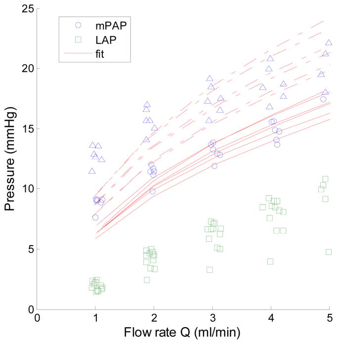
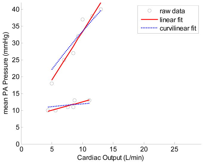
Pulmonary hypertension (PH) is initially a disease of the small, peripheral resistance arteries. Changes in these vessels are best assessed by measurement of pulmonary artery pressure at several levels of flow to generate multi-point pressure-flow curves. This approach is superior to the traditional single-point measurement of pulmonary vascular resistance (PVR) because it allows a flow-independent definition of the resistive properties of that portion of the pulmonary vascular bed and also provides information on its distensibility. In animal models, multi-point pressure-flow curves can be obtained using an isolated, ventilated, perfused lung system. Clinically, cardiopulmonary exercise testing (CPET) with non-invasive echocardiography is feasible and provides realistic values of the resistance and peripheral compliance. Together, these values can be used to better understand and screen for PH and exercise-induced PH.
Figures
Similar articles
-
Extraction of pulmonary vascular compliance, pulmonary vascular resistance, and right ventricular work from single-pressure and Doppler flow measurements in children with pulmonary hypertension: a new method for evaluating reactivity: in vitro and clinical studies.Circulation. 2004 Oct 26;110(17):2609-17. doi: 10.1161/01.CIR.0000146818.60588.40. Epub 2004 Oct 18. Circulation. 2004. PMID: 15492299
-
How to measure pulmonary vascular and right ventricular function.Annu Int Conf IEEE Eng Med Biol Soc. 2009;2009:177-80. doi: 10.1109/IEMBS.2009.5333835. Annu Int Conf IEEE Eng Med Biol Soc. 2009. PMID: 19964469 Free PMC article. Review.
-
Loss of Vascular Distensibility During Exercise Is an Early Hemodynamic Marker of Pulmonary Vascular Disease.Chest. 2016 Feb;149(2):353-361. doi: 10.1378/chest.15-0125. Epub 2016 Jan 12. Chest. 2016. PMID: 26134583
-
Left Atrial Intrinsic Strain Rate Correcting for Pulmonary Wedge Pressure Is Accurate in Estimating Pulmonary Vascular Resistance in Breathless Patients.Echocardiography. 2016 Aug;33(8):1156-65. doi: 10.1111/echo.13226. Epub 2016 May 4. Echocardiography. 2016. PMID: 27144613
-
Exercise physiology and pulmonary arterial hypertension.Prog Cardiovasc Dis. 2012 Sep-Oct;55(2):172-9. doi: 10.1016/j.pcad.2012.07.003. Prog Cardiovasc Dis. 2012. PMID: 23009913 Review.
Cited by
-
Pulmonary vascular wall stiffness: An important contributor to the increased right ventricular afterload with pulmonary hypertension.Pulm Circ. 2011 Apr-Jun;1(2):212-23. doi: 10.4103/2045-8932.83453. Pulm Circ. 2011. PMID: 22034607 Free PMC article.
-
Discrepancy between pulmonary arterial wedge pressure and left ventricular end diastolic pressure in patients with interstitial lung disease.JHLT Open. 2024 Sep 29;6:100160. doi: 10.1016/j.jhlto.2024.100160. eCollection 2024 Nov. JHLT Open. 2024. PMID: 40145039 Free PMC article.
-
Exogenous Estrogen Preserves Distal Pulmonary Arterial Mechanics and Prevents Pulmonary Hypertension in Rats.Am J Respir Crit Care Med. 2020 Feb 1;201(3):371-374. doi: 10.1164/rccm.201906-1217LE. Am J Respir Crit Care Med. 2020. PMID: 31661294 Free PMC article. No abstract available.
-
Pulmonary vascular mechanics: important contributors to the increased right ventricular afterload of pulmonary hypertension.Exp Physiol. 2013 Aug;98(8):1267-73. doi: 10.1113/expphysiol.2012.069096. Epub 2013 May 10. Exp Physiol. 2013. PMID: 23666792 Free PMC article. Review.
-
Measuring right ventricular function in the normal and hypertensive mouse hearts using admittance-derived pressure-volume loops.Am J Physiol Heart Circ Physiol. 2010 Dec;299(6):H2069-75. doi: 10.1152/ajpheart.00805.2010. Epub 2010 Oct 8. Am J Physiol Heart Circ Physiol. 2010. PMID: 20935149 Free PMC article.
References
-
- McLaughlin VV, Archer SL, Badesch DB, et al. ACCF/AHA 2009 Expert Consensus Document on Pulmonary Hypertension. A Report of the American College of Cardiology Foundation Task Force on Expert Consensus Documents and the American Heart Association. Circulation. 2009;119 doi: 10.1161/CIRCULATIONAHA.109.192230. - DOI - PubMed
-
- Reeves JT, Dempsey JA, Grover RF. Pulmonary circulation during exercise. In: Weir EK, Reeves JT, editors. Pulmonary Vascular Physiology and Physiopathology. Vol. 4. Marcel Dekker; New York: 1989. pp. 107–133.
-
- Castelain V, Chemla D, Humbert M, et al. Pulmonary artery pressure flow relations after prostacyclin in primary pulmonary hypertension. Am J Respir Crit Care Med. 2002;165 (3):338–340. - PubMed
-
- Reeves JT, Linehan JH, Stenmark KR. Distensibility of the normal human lung circulation during exercise. Am J Physiol Lung Cell Mol Physiol. 2005;288:L419–25. - PubMed
Publication types
MeSH terms
Grants and funding
LinkOut - more resources
Full Text Sources
Medical
Research Materials