. 2009:2009:2719-22.
doi: 10.1109/IEMBS.2009.5333386.

Classification in DTI using shapes of white matter tracts

Affiliations

Classification in DTI using shapes of white matter tracts

Nagesh Adluru et al. Annu Int Conf IEEE Eng Med Biol Soc. 2009.

Abstract

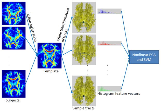
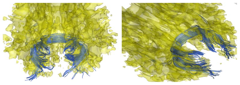
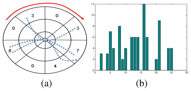
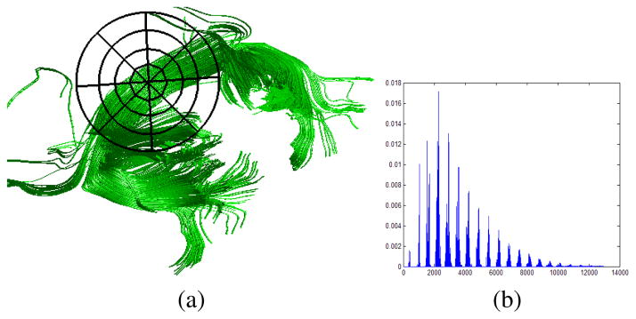
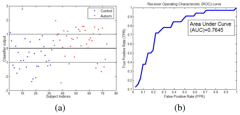
Diffusion Tensor Imaging (DTI) provides unique information about the underlying tissue structure of brain white matter in vivo, including both the geometry of fiber bundles as well as quantitative information about tissue properties as characterized by measures such as tensor orientation, anisotropy, and size. Our objective in this paper is to evaluate the utility of shape representations of white matter tracts extracted from DTI data for classification of clinically different population groups (here autistic vs control). As a first step, our algorithm extracts fiber bundles passing through approximately marked regions of interest on affinely aligned brain volumes. The subsequent analysis is entirely based on the geometric modeling of the extracted tracts. A key advantage of using such an abstraction is that it allows us to capture invariant features of brains allowing for efficient large sample size studies. We demonstrate that with the use of an appropriate representation of the tract shapes, classifiers can be built with reasonable prediction accuracies without making heavy use of the spatial normalization machinery needed when using voxel based features.


Figures

Fig. 1
Fig. 1
Schematic overview of the shape based classification system. Subjects are registered to a template using affine transformations. These affine transformations are used to align the tracts passing through the splenium. The tracts are represented using 3D shape context. These histogram based feature vectors are projected into a low dimensional space using non-linear PCA and classified using binary SVM.
Fig. 2
Fig. 2
Sample tracts (blue) passing through splenium of the corpus callosum for two subjects.
Fig. 3
Fig. 3
Shape context. Demonstration in 2D for ease of understanding. (a) The log-polar binning used in 2D shape context [11]. Sample 2D streamlines are shown as dotted blue curves. Counts for outermost radial bins are also shown. By reading the bins in the order pointed by the red curve in (a) we obtain the histogram in (b).
Fig. 4
Fig. 4
(a) Sample tracts passing through the splenium. A sample 2D version of binning-frame is centered at the splenium. (b) The histogram representation for the tracts.
Fig. 5
Fig. 5
(a) Classifier output (h(x) − b, Eq. (3)) values for the two classes. The thick line is the classification boundary and the dotted lines are the margins. Values above the thick line indicate autism and those below indicate control subjects. Examples inside the margins are harder examples. (b) ROC curve shows that our classifier can perform reasonably well with an area under curve (AUC) of 0.7645. Average specificity and sensitivity is 71.88%. The values are estimated using leave-one-out cross validation.

References

    1. Lazar M, Weinstein D, Tsuruda J, Hasan K, et al. White matter tractography using diffusion tensor deflection. Human Brain Mapping. 2003;18(4) - PMC - PubMed
    1. Savadjiev P, Campbell JS, Pike GB, Siddiqi K. MICCAI. Berlin, Heidelberg: Springer-Verlag; 2008. Streamline flows for white matter fibre pathway segmentation in diffusion MRI; pp. 135–143. - PubMed
    1. King MD, Gadiana DG, Clarka CA. A random effects modelling approach to the crossing-fibre problem in tractography. NeuroImage. 2009 Feb;44(3):753–768. - PubMed
    1. O’Donnel L, Westin CF, Golby A. Tract-based morphometry for white matter group analysis. NeuroImage. 2009;45:832–844. - PMC - PubMed
    1. Goodlett CB, Fletcher TP, Gilmore JH, Gerig G. Group analysis of DTI fiber tract statistics with application to neurodevelopment. NeuroImage. 2009;45(1) - PMC - PubMed

Publication types

LinkOut - more resources