Alignment and elongation of human adipose-derived stem cells in response to direct-current electrical stimulation
- PMID: 19964171
- PMCID: PMC2791914
- DOI: 10.1109/IEMBS.2009.5333142
Alignment and elongation of human adipose-derived stem cells in response to direct-current electrical stimulation
Abstract
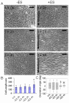
In vivo, direct current electric fields are present during embryonic development and wound healing. In vitro, direct current (DC) electric fields induce directional cell migration and elongation. For the first time, we demonstrate that cultured human adipose tissue-derived stem cells (hASCs) respond to the presence of direct-current electric fields. Cells were stimulated for 2-4 hours with DC electric fields of 6 V/cm that were similar to those encountered in vivo post-injury. Upon stimulation, hASCs were observed to elongate and align perpendicularly to the applied electric field, disassemble gap junctions, and upregulate the expression of genes for connexin-43, thrombomodulin, vascular endothelial growth factor, and fibroblast growth factor. In separate related studies, human epicardial fat-derived stem cells (heASCs) were also observed to align and elongate. It is interesting that the morphological and phenotypic characteristics of mesenchymal stem cells derived both from liposuction aspirates and from cardiac fat can be modulated by direct current electric fields. In further studies, we will quantify the effects of the electrical fields in the context of wound healing.
Figures


Similar articles
-
Osteogenic differentiation of human adipose-derived stem cells can be accelerated by controlling the frequency of continuous ultrasound.J Ultrasound Med. 2013 Aug;32(8):1461-70. doi: 10.7863/ultra.32.8.1461. J Ultrasound Med. 2013. PMID: 23887957
-
[Preliminary evaluation and mechanism of adipose-derived stem cell transplantation from allogenic diabetic rats in the treatment of diabetic rat wounds].Zhonghua Shao Shang Za Zhi. 2019 Sep 20;35(9):645-654. doi: 10.3760/cma.j.issn.1009-2587.2019.09.002. Zhonghua Shao Shang Za Zhi. 2019. PMID: 31594182 Chinese.
-
Low-intensity current (LIC) stimulation of subcutaneous adipose derived stem cells (ADSCs) - A missing link in the course of LIC based wound healing.Med Hypotheses. 2019 Apr;125:79-83. doi: 10.1016/j.mehy.2019.02.039. Epub 2019 Feb 16. Med Hypotheses. 2019. PMID: 30902156
-
The potential of adipose-derived adult stem cells as a source of neuronal progenitor cells.Plast Reconstr Surg. 2005 Oct;116(5):1453-60. doi: 10.1097/01.prs.0000182570.62814.e3. Plast Reconstr Surg. 2005. PMID: 16217495 Review.
-
Our Fat Future: Translating Adipose Stem Cell Therapy.Stem Cells Transl Med. 2015 Sep;4(9):974-9. doi: 10.5966/sctm.2015-0071. Epub 2015 Jul 16. Stem Cells Transl Med. 2015. PMID: 26185256 Free PMC article. Review.
Cited by
-
Surface-patterned electrode bioreactor for electrical stimulation.Lab Chip. 2010 Mar 21;10(6):692-700. doi: 10.1039/b917743d. Epub 2010 Jan 5. Lab Chip. 2010. PMID: 20221556 Free PMC article.
-
Recent advances in soluble decellularized extracellular matrix for heart tissue engineering and organ modeling.J Biomater Appl. 2023 Nov;38(5):577-604. doi: 10.1177/08853282231207216. J Biomater Appl. 2023. PMID: 38006224 Free PMC article. Review.
-
Current progress in use of adipose derived stem cells in peripheral nerve regeneration.World J Stem Cells. 2015 Jan 26;7(1):51-64. doi: 10.4252/wjsc.v7.i1.51. World J Stem Cells. 2015. PMID: 25621105 Free PMC article. Review.
-
Direct electrical stimulation enhances osteogenesis by inducing Bmp2 and Spp1 expressions from macrophages and preosteoblasts.Biotechnol Bioeng. 2019 Dec;116(12):3421-3432. doi: 10.1002/bit.27142. Epub 2019 Sep 23. Biotechnol Bioeng. 2019. PMID: 31429922 Free PMC article.
-
Bioactive polymeric materials and electrical stimulation strategies for musculoskeletal tissue repair and regeneration.Bioact Mater. 2020 Apr 7;5(3):468-485. doi: 10.1016/j.bioactmat.2020.03.010. eCollection 2020 Sep. Bioact Mater. 2020. PMID: 32280836 Free PMC article. Review.
References
-
- Levin M. Motor protein control of ion flux is an early step in embryonic left-right asymmetry. BioEssays. 2003;25:1002–1010. - PubMed
-
- Finkelstein E, Chang W, Chao PHG, Gruber D, Minden A, Hung CT, Bulinski JC. Roles of microtubules, cell polarity and adhesion in electric-field-mediated motility of 3T3 fibroblasts. J Cell Sci. 2004;117(8):1533–1545. - PubMed
-
- Chao P-HG, Roy R, Mauck RL, Liu W, Valhmu WB, Hung CT. Chondrocyte Translocation Response to Direct Current Electric Fields. Journal of Biomechanical Engineering. 2000;122(3):261–267. - PubMed
-
- Ferrier J, Ross SM, Kanehisa J, Aubin JE. Osteoclasts and osteoblasts migrate in opposite directions in response to a constant electrical field. J. Cell. Physiol. 1986;129(3):283–288. - PubMed
-
- Gruler H, Nuccitelli R. Neural crest cell galvanotaxis: new data and a novel approach to the analysis of both galvanotaxis and chemotaxis. Cell Motil. Cytoskeleton. 1991;19(2):121–133. - PubMed
Publication types
MeSH terms
Grants and funding
LinkOut - more resources
Full Text Sources
Other Literature Sources
Medical
Miscellaneous
