

Review
. 2009 Oct 2;9 Spec No A(Special issue A):S38-43.
doi: 10.1102/1470-7330.2009.9009.
The incidental skeletal lesion: ignore or explore?
Affiliations
- PMID: 19965292
- PMCID: PMC2797467
- DOI: 10.1102/1470-7330.2009.9009
Item in Clipboard
Review
The incidental skeletal lesion: ignore or explore?
Cancer Imaging.
.
Abstract
The 'leave me alone' bone lesions are very classical, and, as indicated by their name, do not require any further investigation. There are very typical cases, and there are also more difficult ones, and they can be especially difficult to manage if the patient has a known cancer.
Figures
This child has a nephroblastoma. The lytic painless lesion of the femur has a regular well-demarcated medial border on anteroposterior (AP) radiographs (a) and lateral view (b). The lateral view is poorly limited. That led to a CT scan (c). Bilateral lesions are easily detected. Despite the lack of calcified periosteum, the lesion is typical of non-ossifying fibromas. The lateral border was not sharp on the AP radiograph because of its oblique direction.

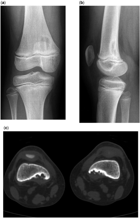
Cortical desmoids: radiographs (a), CT (b), coronal MR (c), histology (d), and radiograph 3 weeks after the biopsy (e). The lytic lesion looks aggressive, with cortical destruction and perpendicular periosteal reactions. The typical location, on the cortex of the metaphysic of the medial codyle was so suggestive of the diagnosis that the biopsy should not have been performed. Histologically, the irregular young bone formation suggested an osteosarcoma. The follow-up film, displaying the calcified hematoma, made the diagnosis still more difficult. Everything went back to normal, without further treatment.

Incidental discovery of a regular, well-limited, ground glass lesion, very typical of a fibrous dysplasia on radiograph (a), CT (b) and MRI (c).

Sprain of the knee while jogging. On radiographs (lateral view; a), a calcified mass typical of cartilage was discovered. On MR (a, sagittal T1; b, sagittal T2FS; c, axial PD), both the bone bruise and the avulsion of the anterior cruciate ligament, and the cartilage tumour were detected. The pain disappeared within 3 weeks. Despite the partial erosion of the cortex, a decision not to treat the probable low grade cartilaginous tumour was made, and the lesion is stable 10 years later.

Renal cell carcinoma and pain in the thigh. On radiographs (a) a cortical lytic lesion was detected and confirmed on CT (b). CT also revealed multiple sclerotic, speculated, bone lesions (c–g) typical of benign bone islands.
References
-
- Yanagawa T, Watanabe H, Shinozaki T, Ahmed AR, Shirakura K, Takagishi K. The natural history of disappearing bone tumours and tumour-like conditions. Clin Radiol. 2001;56:877–86. doi:10.1053/crad.2001.0795. PMid:11603890. - DOI - PubMed
-
- Kontogeorgakos VA, Xenakis T, Papachristou D, et al. Cortical desmoid and the four clinical scenarios. Arch Orthop Trauma Surg. 2009;129:779–85. doi:10.1007/s00402-008-0687-6. PMid:18612646. - DOI - PubMed
-
- Masciocchi C, Sparvoli L, Barile A. Diagnostic imaging of malignant cartilage tumors. Eur J Radiol. 1998;27(Suppl 1):S86–90. doi:10.1016/S0720-048X(98)00048-5. PMid:9652507. - DOI - PubMed
-
- Kyriakos M, McDonald DJ, Sundaram M. Fibrous dysplasia with cartilaginous differentiation (“fibrocartilaginous dysplasia”): a review, with an illustrative case followed for 18 years. Skeletal Radiol. 2004;33:51–62. doi:10.1007/s00256-003-0718-x. PMid:14647989. - DOI - PubMed
-
- Skeletal Lesions Interobserver Correlation among Expert Diagnosticians (SLICED) Study Group. Reliability of histopathologic and radiologic grading of cartilaginous neoplasms in long bones. J Bone Joint Surg Am. 2007;89:2113–23. doi:10.2106/JBJS.F.01530. PMid:17908885. - DOI - PubMed
Publication types
MeSH terms
LinkOut - more resources
Full Text Sources
Medical