. 2009 Dec 15;106(50):21413-8.
doi: 10.1073/pnas.0911935106. Epub 2009 Nov 24.

Studies on expression and function of the TMEM16A calcium-activated chloride channel

Affiliations

Studies on expression and function of the TMEM16A calcium-activated chloride channel

Fen Huang et al. Proc Natl Acad Sci U S A. .

Abstract

Calcium-activated chloride channels (CaCC) with similar hallmark features are present in many cell types and mediate important physiological functions including epithelial secretion, sensory signal transduction, and smooth muscle contraction. Having identified TMEM16A of the transmembrane proteins with unknown function (TMEM) 16 family as a CaCC subunit, we have developed antibodies specific for mouse TMEM16A, as evidenced by the absence of immunoreactivity in TMEM16A knockout mice. Here, we show that TMEM16A is located in the apical membranes of epithelial cells in exocrine glands and trachea. In addition, TMEM16A is expressed in airway smooth muscle cells and the smooth muscle cells of reproductive tracts, the oviduct and ductus epididymis. In the gastrointestinal (GI) tract, TMEM16A is absent from smooth muscle cells, but present in the interstitial cells of Cajal (ICC), the pacemaker cells that control smooth muscle contraction. The physiological importance of TMEM16A is underscored by the diminished rhythmic contraction of gastric smooth muscle from TMEM16A knockout mice. The TMEM16A expression pattern established in this study thus provides a roadmap for the analyses of physiological functions of calcium-activated chloride channels that contain TMEM16A subunits.


Conflict of interest statement

The authors declare no conflict of interest.

Figures

Fig. 1.
Fig. 1.
TMEM16A rabbit polyclonal antibody characterization. (A) TMEM16A antibody specifically recognizes the TMEM16A-GFP fusion protein exogenously expressed in HEK293 cells. Note the membrane localization of TMEM16A-GFP. (B) In TMEM16A wild type mice, the mRNA fragment (995-bp) can be detected by RT-PCR with primers flanking exon12, while a shorter mRNA fragment (838-bp) with exon12 omission can be detected in TMEM16A knockout mouse. (C) The mutantTMEM16A-GFP construct does not yield detectable expression of truncated TMEM16A protein in HEK293 cells. (D) TMEM16A antibody specifically recognizes the endogenous TMEM16A protein in acinar cells of salivary glands and the pancreas of the wild type mouse. No expression was observed in tissue from the TMEM16A knockout mouse. The Bottom reveals the apical localization of TMEM16A in the pancreatic acinar cells by double labeling with the basolateral membrane marker E-cadherin. Blue is the nuclear staining with DAPI.
Fig. 2.
Fig. 2.
TMEM16A is expressed in the airway epithelial cells and smooth muscle cells. (A) Lack of TMEM16A immunoreactivity in the airway of the TMEM16A knockout mouse. (B) Smooth muscle actin (SMA) staining in the airway of the TMEM16A knockout mouse. (C) Overlay of A and B. (D) TMEM16A expression in the airway epithelium (yellow arrow) and smooth muscle cells (pink arrowhead) of the wild type mouse. (E) SMA staining marks the smooth muscle cells in the wild type airway. (F) Overlay of D and E. (G) Higher magnification figure of TMEM16A staining in the wild type airway. (H) Higher magnification figure of SMA staining. (I) Overlay of G and H. Note the expression of TMEM16A in the SMA positive SMCs. Blue is the nuclear staining with DAPI.
Fig. 3.
Fig. 3.
TMEM16A is expressed in the SMCs in the reproductive tract. (A–C) TMEM16A is expressed in the SMCs of the oviduct. (D–F) TMEM16A is expressed in the SMCs of the ductus epididymis. Blue is the nuclear staining with DAPI.
Fig. 4.
Fig. 4.
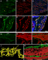
TMEM16A is expressed in the interstitial cells of cajal (ICC) in the mouse stomach and intestine. (A–C) TMEM16A is expressed in C-kit positive ICC in the stomach. Arrows point to examples of spindle shaped ICC-IM. (D–F) TMEM16A is expressed in C-kit positive ICC in the intestine. Arrows point to examples of ICC-SM, ICC-MY, and ICC-IM. (G–I) Double labeling with SMA reveals no detectable expression of TMEM16A in SMCs in the intestine. (J) Three-dimensional surface reconstruction of cells double labeled for TMEM16A and C-kit revealed the multiple processes of ICC-MY and their interconnections forming the ICC network. Small cellular protrusions with detectable immunoreactivity for TMEM16A but not for C-kit can also be seen in the 3-D surface reconstruction. Green is TMEM16A and yellow indicates the colocalization of TMEM16A and C-kit. Pink arrows point out the cell bodies of ICC. (K) Three-dimensional reconstruction illustrates the spatial relationship of TMEM16A-positive ICC and the SMA-positive SMCs in the intestine. Note the ICC-SM on the top of circular smooth muscle, the ICC-MY between the circular and longitudinal smooth muscle layers and the ICC-IM in the longitudinal muscle layer. Green is TMEM16A and red is SMA. Blue is the nuclear staining with DAPI.
Fig. 5.
Fig. 5.
Smooth muscle contraction is diminished in the stomach antrum of the TMEM16A knockout mouse. (A) Representative cell motility along the axis of the circular muscle orientation for the wild type stomach antral smooth muscle. Note the oscillation of the cell position. (B) The cell position variation along the circular muscle axis for the TMEM16A knockout mouse stomach antral smooth muscle is less regular, with significantly reduced frequency and amplitude. (C) Representative trajectories of cell movement in the wild type and knockout mouse stomach. The blue trace is wild type and the red is knockout. X is the circular muscle axis and Y is the longitudinal muscles axis. Note the much larger motility range in the wild type than in the knockout mouse. (D) Quantification of the average total trajectory length of cell motility in the wild type and knockout mouse. The average total trajectory length in the knockout is ≈19.88 ± 0.005% of the wild type (n = 8–10 in three pairs of wild type and knockout siblings).

References

    1. Eggermont J. Calcium-activated chloride channels: (Un)known, (un)loved? Proc Am Thorac Soc. 2004;1:22–27. - PubMed
    1. Hartzell C, Putzier I, Arreola J. Calcium-activated chloride channels. Annu Rev Physiol. 2005;67:719–758. - PubMed
    1. Yang YD, et al. TMEM16A confers receptor-activated calcium-dependent chloride conductance. Nature. 2008;455:1210–1215. - PubMed
    1. Schroeder BC, Cheng T, Jan YN, Jan LY. Expression cloning of TMEM16A as a calcium-activated chloride channel subunit. Cell. 2008;134:1019–1029. - PMC - PubMed
    1. Caputo A, et al. TMEM16A, a membrane protein associated with calcium-dependent chloride channel activity. Science. 2008;322:590–594. - PubMed

Publication types

LinkOut - more resources