. 2009 Dec 18;326(5960):1698-701.
doi: 10.1126/science.1182372. Epub 2009 Nov 12.

The Fanconi anemia pathway promotes replication-dependent DNA interstrand cross-link repair

Affiliations

The Fanconi anemia pathway promotes replication-dependent DNA interstrand cross-link repair

Puck Knipscheer et al. Science. .

Abstract

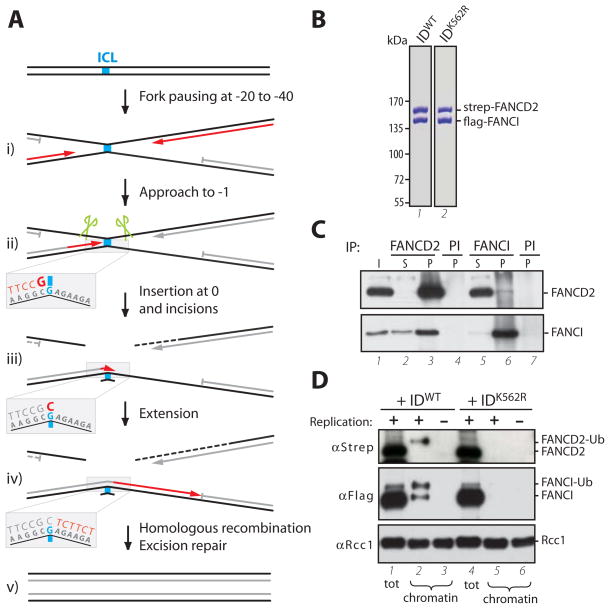
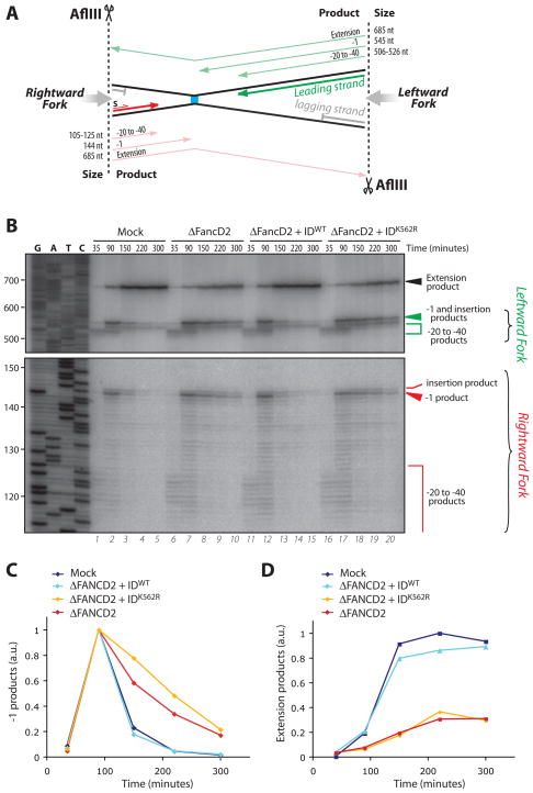
Fanconi anemia is a human cancer predisposition syndrome caused by mutations in 13 Fanc genes. The disorder is characterized by genomic instability and cellular hypersensitivity to chemicals that generate DNA interstrand cross-links (ICLs). A central event in the activation of the Fanconi anemia pathway is the mono-ubiquitylation of the FANCI-FANCD2 complex, but how this complex confers ICL resistance remains enigmatic. Using a cell-free system, we showed that FANCI-FANCD2 is required for replication-coupled ICL repair in S phase. Removal of FANCD2 from extracts inhibits both nucleolytic incisions near the ICL and translesion DNA synthesis past the lesion. Reversal of these defects requires ubiquitylated FANCI-FANCD2. Our results show that multiple steps of the essential S-phase ICL repair mechanism fail when the Fanconi anemia pathway is compromised.


Figures

Fig. 1
Fig. 1
(A) Schematic representation of lesion bypass in ICL repair (12). (B) Purified FANCI-FANCD2WT and FANCI-FANCD2K562R stained with Coomassie blue. (C) Reciprocal co-immunoprecipitation of xlFANCI and xlFANCD2 from Xenopus egg extract. Input (I) and supernatant (S) (0.2 μl extract), or precipitated proteins (P, from 1 μl extract) were blotted for FANCI and FANCD2. PI: pre-Immune serum. (D) Replication-dependent binding of FANCI-FANCD2 to damaged chromatin. Crosslinked sperm chromatin was replicated in undepleted extracts supplemented with FANCI-FANCD2WT or FANCI-FANCD2K562R (310 nM). Chromatin-bound fractions (from 2 μl extract) or total extract (0.2 μl), were analyzed by Western blotting with anti-strep-tag (to visualize recombinant FANCD2), anti-FLAG-tag (recombinant FANCI), and anti-RCC1 (loading control) antibodies. Where indicated, replication was inhibited with Geminin. Note that only ubiquitylated FANCD2 binds chromatin, while both ubiquitylated and unubiquitylated FANCI bind (see also (14)).
Fig. 2
Fig. 2
FANCD2 and its ubiquitylation are required for ICL repair. (A) Sequence surrounding the ICL and relevant restriction sites of pICL. (B) pICL (2.3 ng/μl) was replicated in mock-depleted extract, FANCD2-depleted extract (ΔFANCD2) or ΔFANCD2 extract supplemented with 375 nM FANCI-FANCD2WT or FANCI-FANCD2K562R, and repair efficiency was plotted. For primary data and calculation of repair efficiency, see Fig. S5A.
Fig. 3
Fig. 3
Insertion of a nucleotide across from the damaged base is compromised in FANCD2-depleted extracts (A) Schematic representation of leading strand intermediates generated after pICL digestion with AflIII. (B) Samples from the reactions described in Fig. 2B were digested with AflIII, separated on a sequencing gel alongside a ladder derived from extension of primer S (S, in panel A) on pControl, and visualized via autoradiography. Nascent strands generated by the rightward (red) and leftward (green) replication forks are indicated to the right and illustrated in (A). −1 and extension products observed in (B) were quantified and graphed in (C) and (D), respectively.
Fig. 4
Fig. 4
ICL-proximal incisions are compromised in FANCD2-depleted extracts. (A) Schematic representation of predicted fragments generated by HincII digestion of pICL, before and after dual incisions and lesion bypass. Parental strands in black, nascent strands in grey. (B) pICL (2.5 ng/μl) was replicated in mock-depleted extract, FANCD2-depleted extract (ΔFANCD2) alone or supplemented with 386 nM FANCI-FANCD2WT. Samples were digested with HincII and separated on a denaturing agarose gel. Both parental and nascent strands are detected by Southern blotting. Unreplicated pControl and pICL were optionally digested with the indicated enzymes and used as size markers for arm/linear species and X-shaped molecules, respectively (lanes 1–3). Note the small amount of linear products in lane 3 (3% of the total), which represents contaminating non-crosslinked plasmids. X-structures and linear species observed in (B) were quantified and graphed in (C) and (D), respectively.

Similar articles

Cited by

References

    1. D’Andrea AD, Grompe M. Nat Rev Cancer. 2003 Jan;3:23. - PubMed
    1. Patel KJ, Joenje H. DNA Repair (Amst) 2007 Jul 1;6:885. - PubMed
    1. Alpi AF, Pace PE, Babu MM, Patel KJ. Mol Cell. 2008 Dec 26;32:767. - PubMed
    1. Garcia-Higuera I, et al. Mol Cell. 2001 Feb;7:249. - PubMed
    1. Smogorzewska A, et al. Cell. 2007 Apr 20;129:289. - PMC - PubMed

Publication types

MeSH terms

Associated data