Review
. 2009 Nov 27;326(5957):1208-12.
doi: 10.1126/science.1175862.

Actin, a central player in cell shape and movement

Affiliations
Review

Actin, a central player in cell shape and movement

Thomas D Pollard et al. Science. .

Abstract

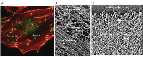
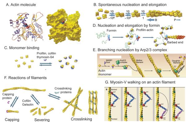
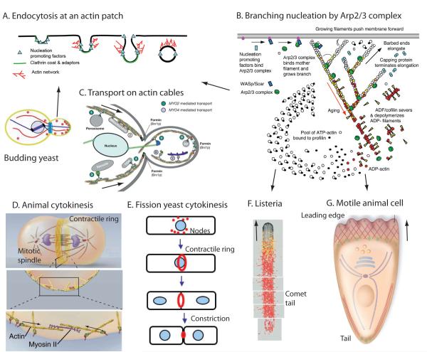
The protein actin forms filaments that provide cells with mechanical support and driving forces for movement. Actin contributes to biological processes such as sensing environmental forces, internalizing membrane vesicles, moving over surfaces, and dividing the cell in two. These cellular activities are complex; they depend on interactions of actin monomers and filaments with numerous other proteins. Here, we present a summary of the key questions in the field and suggest how those questions might be answered. Understanding actin-based biological phenomena will depend on identifying the participating molecules and defining their molecular mechanisms. Comparisons of quantitative measurements of reactions in live cells with computer simulations of mathematical models will also help generate meaningful insights.


Figures

Figure 1
Figure 1
Micrographs of actin filament structures in cells. A, Fluorescent light micrograph of an animal epithelial cell grown in tissue culture and infected with a bacterium, Listeria monocytogenese. Actin filaments are red and bacteria are green. Actin bundles, called stress fibers, bridge sites of adhesion to the substrate. The bacteria use Arp2/3 complex to assemble comet tails for transport through the cytoplasm. B, Electron micrograph of three types of cytoskeletal polymers in a cell permeabilized to release soluble components. After rapid freezing, the frozen water was sublimed away and cellular components were coated with platinum. Red colorization highlights a microtubule. Bundle of actin filaments and a network of intermediate filaments are labeled. C. Electron micrograph of the network of branched actin filaments at the leading edge (top) of a motile keratocyte. The cell was grown in tissue culture, extracted to release soluble materials, dried and coated with platinum. (All images from T.D. Pollard and W.C. Earnshaw, Cell Biology, W.B. Saunders, 2007. Sources are (A) Matthew Welch, (B) John Heuser and (C) Tatyana Svitkina and Gary Borisy.)
Figure 2
Figure 2
Structures of actin and diagrams of fundamental reactions. A, Ribbon and space filling models of the actin molecule (pdb:1ATN). B, Spontaneous nucleation and elongation. Dimers and trimers are unstable. Longer polymers grow rapidly at the barbed end (B) and slowly at the pointed end (P). C, Actin monomer binding proteins. Thymosin-ß1 blocks all assembly reactions; profilin promotes nucleotide exchange and inhibits pointed end elongation and nucleation but not barbed end elongation; cofilin inhibits nucleotide exchange and promotes nucleation. D, Nucleation and elongation by formins. Formins initiate polymerization from free actin monomers and remain associated with the growing barbed end. Profilin-actin binds to formin and transfers actin onto the barbed end of the filament. E, Nucleation by Arp2/3 complex. Nucleation promoting factors such as WASp bind an actin monomer and Arp2/3 complex. Binding to the side of a filament completes activation, and the barbed end of the daughter filament grows from Arp2/3 complex. F, Reactions of actin filaments. Capping proteins bind to and block barbed ends; cofilin and gelsolin sever filaments; crosslinking proteins assemble networks and bundles of actin filaments. G, Myosin motors, such as myosin-V, use cycles of ATP hydrolysis to walk along actin filaments, generally toward the barbed end. (Redrawn from images in T.D. Pollard and W.C. Earnshaw, Cell Biology, W.B. Saunders, 2007.)
Figure 3
Figure 3
Cartoons showing actin based movements. (A) Clathrin-mediated endocytosis at fungal actin patches. (B) Formation of branched filament networks nucleated by Arp2/3 complex, which is used for three of these types of movement, those depicted in panels A, F and G. (C) Transport of membrane-bound vesicles, organelles and RNAs from the mother cell to the daughter cell by class V myosins in budding yeast. (D) Cytokinesis in animal cells by constriction of a contractile ring of actin filaments and myosin-II. (E) Cytokinesis in fission yeast. The contractile ring of actin filaments and myosin-II forms by condensation of nodes. (F) The Listeria bacterium stimulates the assembly of an actin filament comet tail to push it through the cytoplasm of a host animal cell. (G) Locomotion of an animal cell by assembly of actin filaments at the leading edge and retraction of the tail. (A, Redrawn from Ref. 13. B, From Ref. 32. C, Redrawn from Ref. 25. D, Redrawn from T.D. Pollard and W.C. Earnshaw, Cell Biology, W.B. Saunders, 2007. E, From T.D. Pollard. F, Redrawn from Ref. 18. G, Redrawn from Ref. 30.)

Similar articles

Cited by

References

    1. Erickson HP. Bioessays. 2007;29:668. - PMC - PubMed
    1. Richards TA, Cavalier-Smith T. Nature. 2005;436:1113. - PubMed
    1. Geeves MA, Holmes KC. Adv Protein Chem. 2005;71:161. - PubMed
    1. Hatano S, Oosawa F. Biochim. Biophys. Acta. 1966;127:488. - PubMed
    1. Adelman MR, Taylor EW. Biochemistry. 1969 Dec;8:4964. - PubMed

Publication types