Review
. 2009 Nov 27;326(5957):1216-9.
doi: 10.1126/science.1176009.

The extracellular matrix: not just pretty fibrils

Affiliations
Review

The extracellular matrix: not just pretty fibrils

Richard O Hynes. Science. .

Abstract

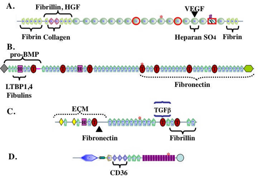
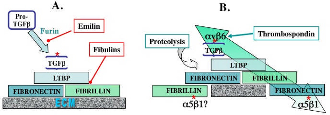
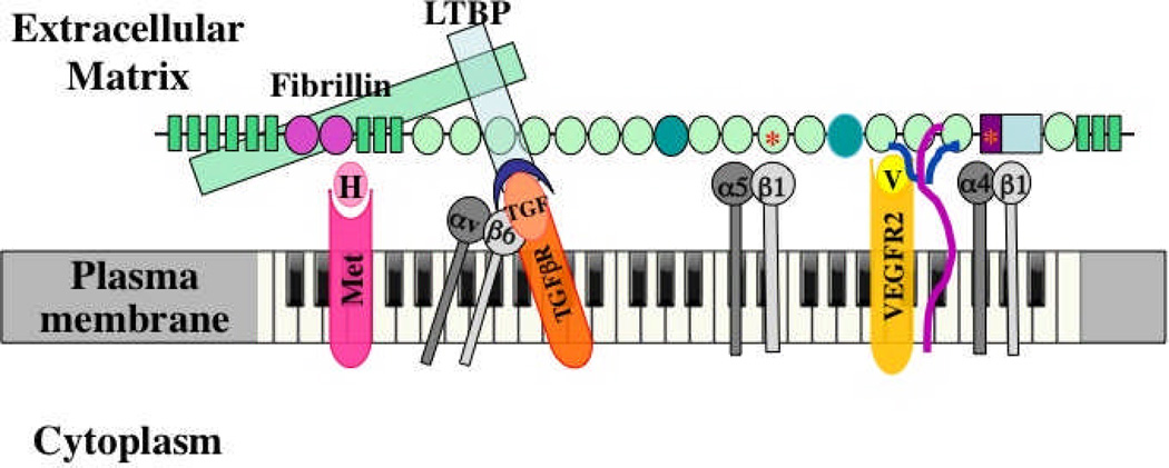
The extracellular matrix (ECM) and ECM proteins are important in phenomena as diverse as developmental patterning, stem cell niches, cancer, and genetic diseases. The ECM has many effects beyond providing structural support. ECM proteins typically include multiple, independently folded domains whose sequences and arrangement are highly conserved. Some of these domains bind adhesion receptors such as integrins that mediate cell-matrix adhesion and also transduce signals into cells. However, ECM proteins also bind soluble growth factors and regulate their distribution, activation, and presentation to cells. As organized, solid-phase ligands, ECM proteins can integrate complex, multivalent signals to cells in a spatially patterned and regulated fashion. These properties need to be incorporated into considerations of the functions of the ECM.


Figures

Figure 1
Figure 1. The complex domain structures of ECM proteins
The figure shows representative ECM proteins (out of hundreds encoded in the genome). The proteins are built from multiple, independently folded domains, which occur in different combinations in different ECM proteins as a consequence of exon shuffling during evolution. The domain structures were generated from the SMART web site (http://smart.embl-heidelberg.de/) and edited in light of specific knowledge about individual proteins. A. Fibronectin. Encoded by a single gene but alternatively spliced at three regions (boxed in red) to generate 12 proteins in rodents and 20 in humans. FN3 domains are widespread in ECM proteins. Binding sites for other matrix proteins are marked. The heparan-sulfate-binding site can interact with proteoglyans (PGs) or with syndecan, an integral membrane PG. The RGD (arg-gly-asp) integrin-binding site is marked by a red asterisk and a second LDV (leu-asp-val) integrin-binding site is marked by a pound sign. Fibronectin is a proangiogenic molecule, whose function is compromised by elimination of the RGD site or of the two alternatively spliced FN3 domains,. FN also binds the proangiogenic growth factors VEGF and HGF,. B. Fibrillin-1. A member of a three-gene family. Fibrillins are made up of EGF-like domains, which are found in many ECM proteins, as well as TB (TGFβ-binding, marked by T) and hybrid (H) domains that are both specific to fibrillins and LTBPs,. Known binding sites within fibrillin-1 for other matrix proteins and growth factors are marked. C. LTBP-1. A member of a four-gene family with structure related to that of fibrillins. Known binding sites for TGFβ/LAP latent complex and for fibrillin and fibronectin are marked. D. Thrombospondin-1. Member of a five-gene family. Thrombospondins 1 and 2 have the structure shown and both are antiangiogenic. Antiangiogenic activity lies in the TSP1 repeats, which bind to the CD36 receptor. TSP1 repeats are also found in other ECM proteins. Thrombospondins also contain EGF-like repeats and a VWC domain, known in other proteins to bind BMPs. The 13 TSP3 repeats (purple) and C-terminal domain are unique to thrombospondins and bind multiple Ca++ ions. In all proteins, the asterisks mark RGD (arg-gly-asp) tripeptide sequences that may bind to integrins, as is well documented for the similar motif in fibronectin (see A).
Figure 2
Figure 2. ECM interactions regulating TGFβ
A. Incorporation into the ECM. Cleavage by furin protease of Pro-TGFβ to the small latent complex (SLC) comprising TGFβ and LAP is inhibited by emilin, an ECM protein. The SLC binds to LTBP, via S-S bonding to a TB domain, to form the large latent complex (LLC), in which form the TGFβ is inactive,. LTBP then binds to fibrillin and to fibronectin (see Figure 1 for specific interaction domains). Fibulins compete for LTBP binding to fibrillin. Fibrillin binds to preexisting fibronectin fibrils or assembles into microfibrils and both fibrillin and fibronectin undergo further homomeric and heteromeric interactions within the ECM. B. Activation of ECM-bound latent TGFβ. TGFβ can be activated by proteolysis of the ECM proteins and/or of LAP or directly by thrombospondin (see text). TGFβ can also be activated by mechanical strain (large green arrow). This strain arises from cytoskeletal force applied through αvβ6 integrin, which binds to an RGD site in LAP and requires attachment of the TGFβ/LAP complex through LTBP to the fibronectin-rich matrix, which is, in turn, attached via α5β1 integrin to other cells. Fibrillin might also be attached to cells via integrins.
Figure 3
Figure 3. Multidomain interactions of ECM proteins with cells
The example shown is fibronectin. Multiple domains are known to bind to integrins, to other ECM proteins and to growth factors, as shown. Integrins α5β1 and α4β1 bind, respectively, to RGD and LDV motifs; heparan sulfate chains of syndecan (purple/blue) bind to FN3-13 as does VEGF. Evidence suggests that VEGF (V, yellow) signals through its own receptor (VEGFR2) more effectively when bound to fibronectin. The same is proposed here for HGF (H) and its receptor (Met, pink). As shown in Figures 1 and 2, fibrillin (green) binds to an N-terminal region of fibronectin and in turn binds LTBP (blue), which recruits TGFβ in a latent complex with LAP (blue crescent). αvβ6 integrin can bind an RGD site in LAP, activating TGFβ, so that it can bind its own receptors (orange). The proposal is that fibronectin organizes and integrates all these signals at two levels. First, by recruiting growth factors to the ECM, fibronectin localizes those signals at the cellular level. Second, the close juxtaposition of the domains in fibronectin brings the different receptors together into an organized submicron patch in the cell surface membrane. Each domain is 2–4 nm in diameter and the entire fibronectin subunit shown is 60–70 nm long, so the receptors will be brought into close apposition such that their signals provide complex, integrated information to the cell – metaphorically generating a melodies and chords in contrast with the “single notes” generated by each receptor. Fibronectin is essential for angiogenesis, and most of the bound receptors and ligands have been shown to play roles in angiogenesis. This model suggests that fibronectin and its associated ECM proteins orchestrate and integrate these signals. In addition, alternatively spliced domains of fibronectin (darker green ovals) are also necessary for proper vascular development and it is a reasonable hypothesis that they introduce additional ligands and/or receptors into the mix.

References

    1. Hynes RO. Cell. 2002;110:673. - PubMed
    1. Berrier AL, Yamada KM. J. Cell Physiol. 2007;213:565. - PubMed
    1. Legate KR, Wickstrom SA, Fassler R. Genes Dev. 2009;23:397. - PubMed
    1. Discher DE, Mooney DJ, Zandstra PW. Science. 2009;324:1673. - PMC - PubMed
    1. Geiger B, Spatz JP, Bershadsky AD. Nat. Rev. Mol. Cell Biol. 2009;10:21. - PubMed

Publication types

MeSH terms

Substances