

Review
doi: 10.1126/science.1175668.
Cell signaling in space and time: where proteins come together and when they're apart
Affiliations
- PMID: 19965465
- PMCID: PMC3041271
- DOI: 10.1126/science.1175668
Item in Clipboard
Review
Cell signaling in space and time: where proteins come together and when they're apart
Science.
.
Abstract
Signal transduction can be defined as the coordinated relay of messages derived from extracellular cues to intracellular effectors. More simply put, information received on the cell surface is processed across the plasma membrane and transmitted to intracellular targets. This requires that the activators, effectors, enzymes, and substrates that respond to cellular signals come together when they need to.
Figures
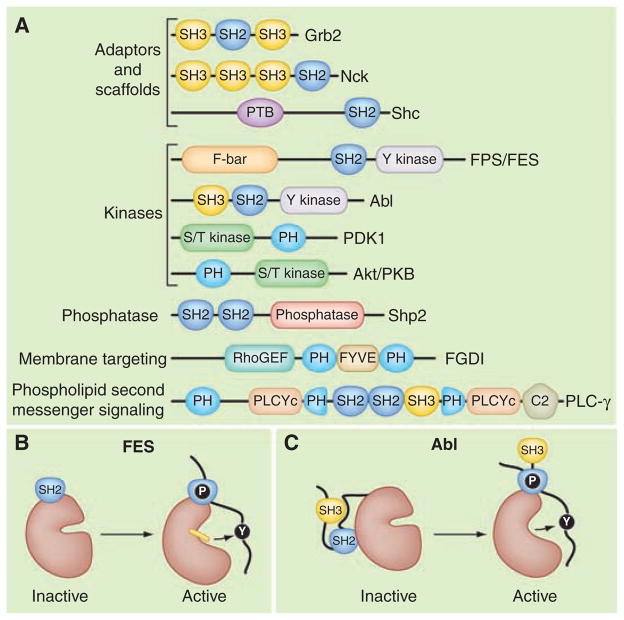
The modular organization of signaling proteins. (A) Schematic diagram depicting the modular domain organization of selected adaptor and scaffolding proteins (Grb2, Nck, Shc); the tyrosine kinases (Fps/FES and Abl); the serine/threonine (S/T) kinases (PDK 1 and Akt/PKB); the protein tyrosine phosphatase (Shp2); a membrane-targeted Rho guanine nucleotide exchange factor (FGDI); and phospholipase C–gamma (PLC-γ). Individual protein modules are indicated: Src homology 2 (SH2); Src homology 3 (SH3); phosphotyrosine binding (PTB); FCHo2-Bin/Amphiphysin/Rvs domain (F-BAR); Fab-1, YGL023, Vps27, and EEA1 domain (FYVE); and Ca2+− dependent membrane-targeting (C2) module. Enzymatic units in each protein are named. (B) Model depicting the active conformation of FES where the SH2 and tyrosine kinase domains form a single functional unit bound to a primed substrate (8). (C) Abl is maintained in an inactive state through the docking of the SH2 domain on the back face of the catalytic core. These intramolecular interactions are broken upon substrate binding (11).
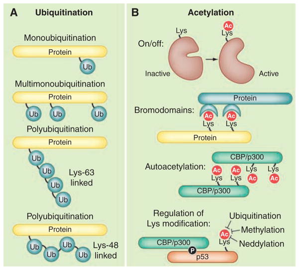
Regulation by ubiquitination or acetylation. These covalent modifications are frequently used for cell communication. (A) Ligation of ubiquitin (Ub) can occur in different patterns on proteins. (B) Acetylation in different contexts serves as an enzymatic on/off switch; for recruitment of bromodomain proteins; (autoacetylation) to induce protein-protein interactions; or for regulation of further lysine modifications.
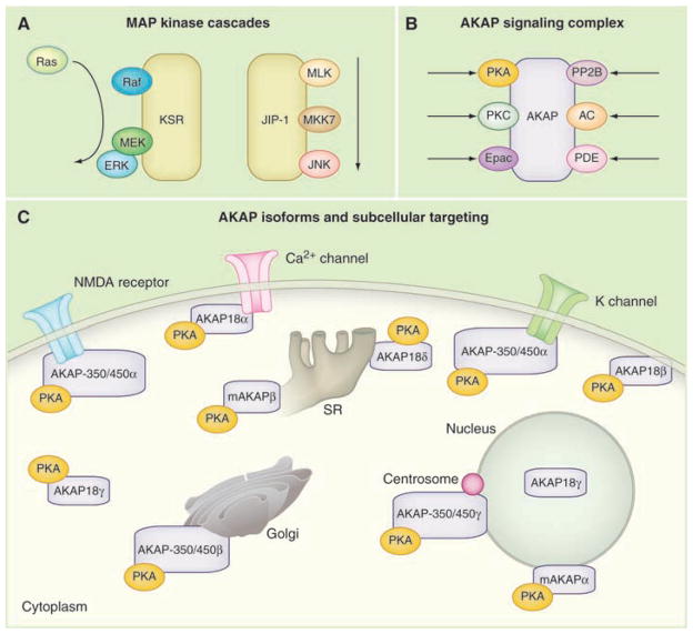
Signal transduction through preassembled enzyme complexes. (A) The linear flow of information through the MAP kinase cascades organized by kinase suppressor of Ras (KSR) and JNK interacting protein (JIP). (B) A-kinase anchoring proteins (AKAPs) organize protein kinase A (PKA), guanine nucleotide exchange factors (Epac) and phosphodiesterases (PDE) into cAMP-responsive complexes. Anchoring of certain protein phosphatases (PP2B) and PKC broaden AKAP function. (C) PKA anchoring to various AKAP isoforms targets the kinase to defined subcellular locations. Diagram of a prototypic cell showing the targeting of PKA via AKAP18, AKAP350/450, and mAKAP variants. NMDA, N-methyl-D-aspartate.
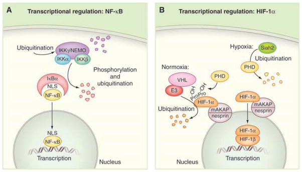
Temporal regulation of signaling events. (A) Schematic diagram of the NF-κB pathway; individual protein components are indicated. (B) The concerted actions of oxygen-sensing enzymes and ubiquitin E3 ligases control the stability of the hypoxia-inducible factor protein HIF-1α. Schematic diagram of the HIF-1α pathway under normoxic (left) and hypoxic (right) conditions; individual protein components are indicated.
References
Publication types
MeSH terms
Substances
Grants and funding
LinkOut - more resources
Full Text Sources
Other Literature Sources
