. 2009 Nov 13;326(5955):948-52.
doi: 10.1126/science.1170633.

How telomeres solve the end-protection problem

Affiliations

How telomeres solve the end-protection problem

Titia de Lange. Science. .

Abstract

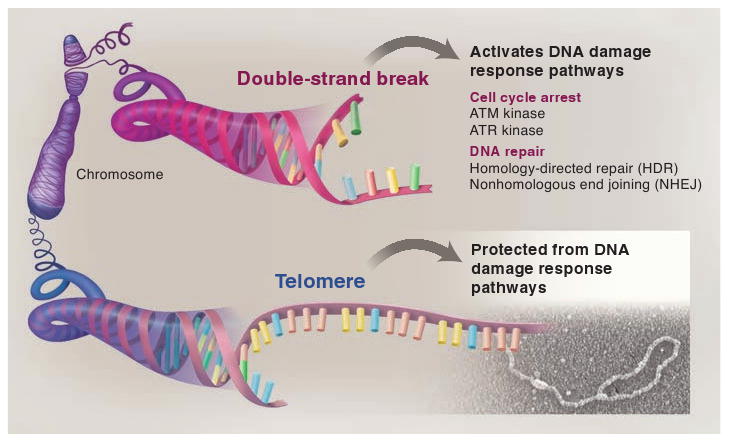
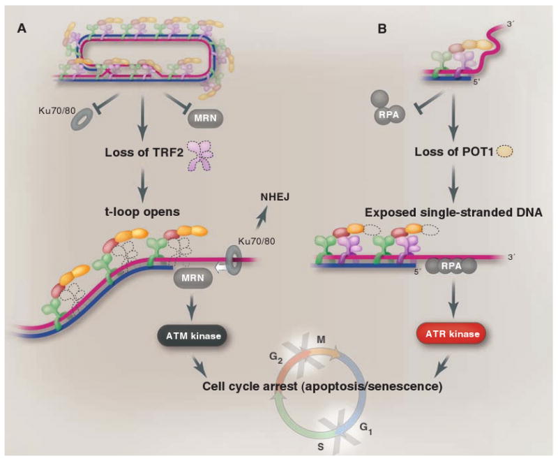
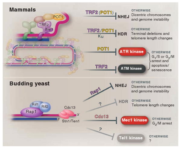
The ends of eukaryotic chromosomes have the potential to be mistaken for damaged or broken DNA and must therefore be protected from cellular DNA damage response pathways. Otherwise, cells might permanently arrest in the cell cycle, and attempts to "repair" the chromosome ends would have devastating consequences for genome integrity. This end-protection problem is solved by protein-DNA complexes called telomeres. Studies of mammalian cells have recently uncovered the mechanism by which telomeres disguise the chromosome ends. Comparison to unicellular eukaryotes reveals key differences in the DNA damage response systems that inadvertently threaten chromosome ends. Telomeres appear to be tailored to these variations, explaining their variable structure and composition.


Figures

Fig. 1
Fig. 1
The end-protection problem. When a mammalian chromosome breaks (top), the exposed DNA ends can activate two signaling pathways (the ATM and ATR kinase pathways) that arrest the cell division cycle and can induce cell death. The broken chromosome is usually repaired by one of two different DNA repair pathways (NHEJ and HDR), allowing cells to continue their divisions with an intact genome. The presence of these DNA damage response pathways poses a problem for the ends of linear chromosomes (telomeres, bottom) because activation of DNA damage signaling or DNA repair at telomeres would be disastrous. Mammalian telomeres solve this end-protection problem through the use of a telomere-specific protein complex (shelterin) and an altered structure (the t-loop) that together ensure that all four pathways remain blocked.
Fig. 2
Fig. 2
Mammalian telomeres. Human and mouse telomeres are composed of long stretches of the repetitive sequence TTAGGG and a telomere-specific protein complex, shelterin (upper left). Shelterin derives its specificity for telomeric DNA from three DNA binding proteins (lower left). TRF1 and TRF2 are two similar proteins that bind to the double-stranded telomeric repeats while POT1 interacts with TTAGGG repeats in single-stranded form. TIN2 and TPP1 connect POT1 to TRF1 and TRF2. Rap1 is bound to TRF2. Telomeres are found in a lariat conformation (upper right), the t-loop, which results from the strand invasion of the 3′ single-stranded overhang into the double-stranded telomeric DNA. Shelterin is sufficiently abundant to cover most of the double-stranded telomeric DNA, and there is sufficient POT1 to cover single-stranded telomeric DNA either in the 3′ overhang or in the D loop. Telomeres also contain nucleosomes and numerous shelterin-associated proteins (not shown).
Fig. 3
Fig. 3
Different components of shelterin are dedicated to different aspects of the end-protection problem. TRF2 represses the ATM kinase signaling pathway (A), whereas POT1 ensures that the ATR kinase is not activated (B). In addition, TRF2 is the main repressor of NHEJ at telomeres, although POT1 contributes to the repression of NHEJ, especially after DNA replication. Both TRF2 and POT1 function to block HDR at telomeres (not shown). TRF2 is proposed to block NHEJ and ATM kinase signaling by forming the t-loop. In the t-loop structure (A), the telomere end is hidden from the DNA end sensor MRN that activates the ATM kinase pathway (MRN), and the Ku70/80 ring (which initiates NHEJ) will not be able to load onto the chromosome end. In (B), POT1 is proposed to block ATR kinase signaling by preventing the binding of RPA, the single-stranded DNA binding protein that activates the ATR kinase pathway. POT1 could block RPA from binding to the single-stranded telomeric DNA either when present at the telomere terminus (as shown) or when exposed in the D loop of the t-loop configuration.
Fig. 4
Fig. 4
Different solutions to the end-protection problem. At mammalian telomeres, the presence of shelterin and the t-loop structure together ensure the repression of the four pathways that threaten telomeres throughout the cell cycle (top). The DNA damage response in budding yeast is not the same as in mammalian cells, hence budding yeast telomeres face a different set of threats (bottom). Whereas Mec1 (ATR equivalent) is a major threat, Tel1 (ATM-like) is not, and HDR is less stringently repressed at budding yeast telomeres than in mammals. Budding yeast telomeres appear tailored to cope with this simpler set of problems, which may explain why none of the shelterin components, except for Rap1, are conserved (bottom). Shelterin is at telomeres throughout the cell cycle, whereas Cdc13/Stn1/Ten1 is not at telomeres before the initiation of DNA replication (not shown).

Similar articles

Cited by

References

    1. Szostak JW, Blackburn EH. Cell. 1982;29:245. - PubMed
    1. Shampay J, Szostak JW, Blackburn EH. Nature. 1984;310:154. - PubMed
    1. Greider CW, Blackburn EH. Cell. 1985;43:405. - PubMed
    1. de Lange T. Nat Rev Mol Cell Biol. 2004;5:323. - PubMed
    1. Orr-Weaver TL, Szostak JW, Rothstein RJ. Proc Natl Acad Sci USA. 1981;78:6354. - PMC - PubMed

Publication types

MeSH terms