. 2010 May;51(5):923-32.
doi: 10.1194/jlr.M000059. Epub 2009 Nov 5.

Novel 14,21-dihydroxy-docosahexaenoic acids: structures, formation pathways, and enhancement of wound healing

Affiliations

Novel 14,21-dihydroxy-docosahexaenoic acids: structures, formation pathways, and enhancement of wound healing

Yan Lu et al. J Lipid Res. 2010 May.

Abstract

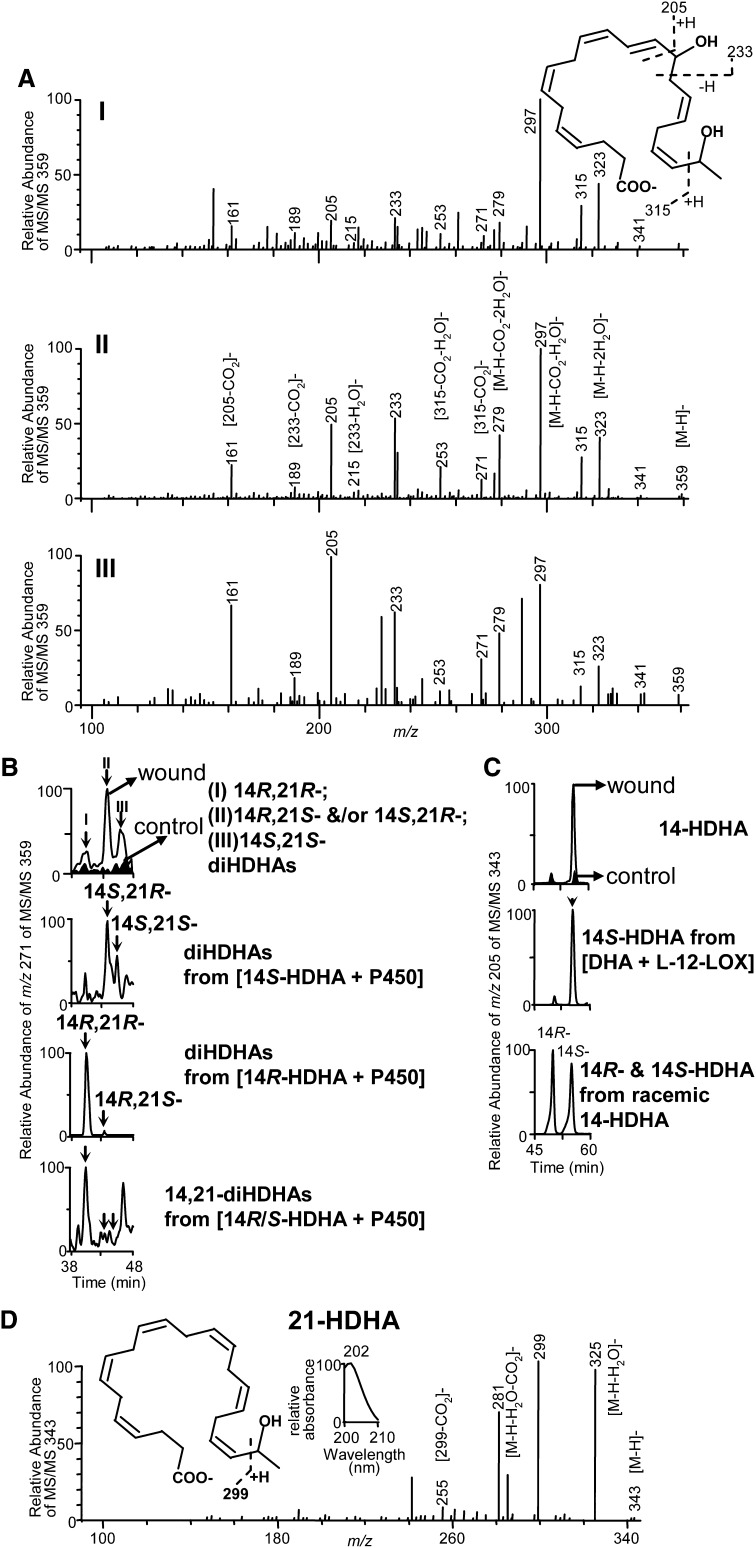
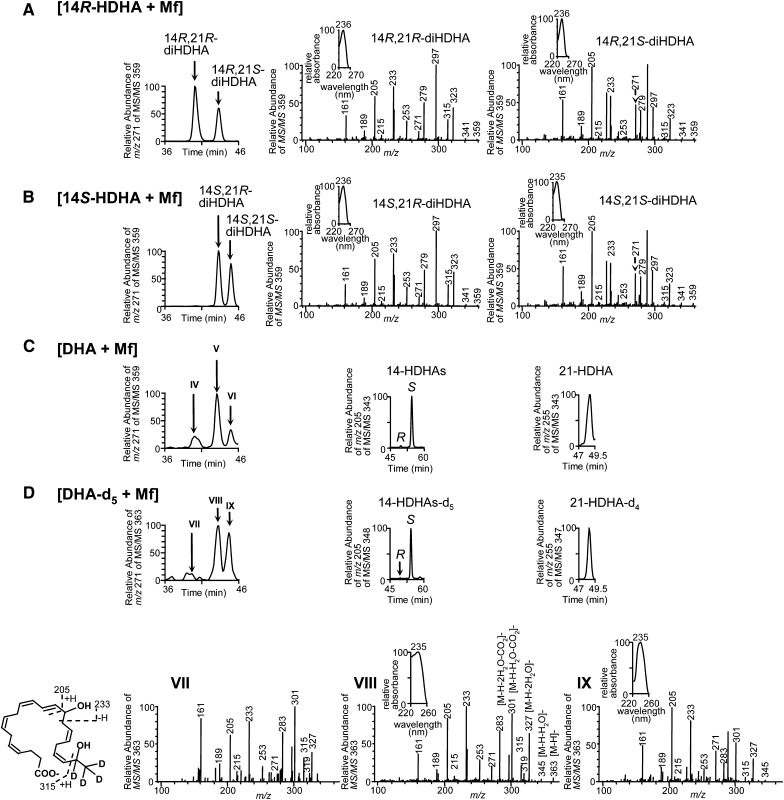
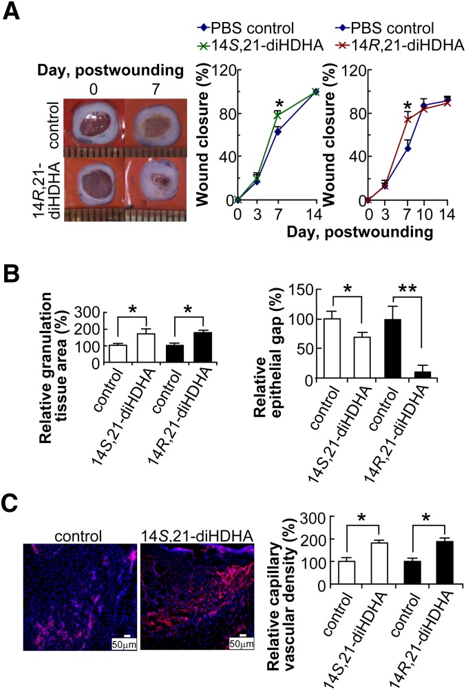
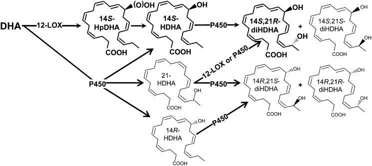
Chronic wounds remain a medical challenge, where well-coordinated cellular and molecular processes required by optimal healing are impaired by diabetes, aging, or other diseases. In determining mechanisms that regulate wound healing, we found that wounding induced formation of novel endogenous 14S,21S-dihydroxy-docosa-4Z,7Z,10Z,12E,16Z,19Z-hexaenoic acids (14S,21S-diHDHA);14R,21R-diHDHA; 14S,21R-diHDHA; and/or 14R,21S-diHDHA. 12-lipoxygenase and cytochrome P450 catalysis in tandem converted docosahexaenoic acid to 14S,21R-diHDHA and 14S,21S-diHDHA through the intermediacy of 14S-HDHA; P450 also converted 14R-HDHA to novel 14R,21R-diHDHA and 14R,21S-diHDHA. Macrophages function as the combination of 12-lipoxgenase and P450 to generate these 14,21-diHDHA stereoisomers, as well as their intermediates 14S-HDHA, 14R-HDHA, and 21-HDHA. The structure and formation pathways of 14,21-diHDHA stereoisomers were further confirmed by macrophage biosynthesis of 14,21-diHDHA-21,22,22,22-d(4) stereoisomers, 14S-HDHA-d(5), 14R-HDHA-d(5), and 21-HDHA-d(4) from DHA-21,21,22,22,22-d(5). We found that 14S,21-diHDHA and 14R,21-diHDHA enhanced wound closure, reepithelialization, granulation tissue growth, and capillary vasculature formation of murine wounds. 14S,21-diHDHA and 14R,21-diHDHA produced by macrophages may partially represent the molecular mechanisms for macrophage pro-healing function. Taken together, 14,21-dihydroxy-DHA stereoisomers and their formation pathways may represent a novel mechanism in the orchestration of wound healing processes, which may provide new insight for developing novel therapeutic modalities that counteract impairments to wound healing.


Figures

Fig. 1.
Fig. 1.
Formation of endogenous 14S,21-diHDHAs and 14R,21-diHDHAs is induced by wounding: molecular structures and formation pathways. Wound tissues were collected from Balb/c mice 3 d after the splinted excisional wounding was performed (n = 3). A: Three typical spectra of chiral LC-MS/MS chromatographic peaks I, II, and III (see 1B) demonstrate the 14,21-diHDHA structure (inset) where the diagnostic MS/MS ions are illustrated. B: Chiral-LC-MS/MS chromatograms show that wounding induced endogenous 14,21-diHDHAs are 14R,21R-diHDHA (I), 14S,21R-diHDHA or 14R,21S-diDHA (II), and 14S,21S-diHDHA (III) which were identified through matching their chiral LC-MS/MS chromatograms and spectra to those of P450-generated 14S-HDHA-derived 14S,21R-diHDHA and 14S,21S-diHDHA (upper middle) as well as 14R-HDHA-derived 14R,21R-diHDHA and 14R,21S-diHDHA (lower middle). Bottom panel is the chiral LC-MS/MS chromatogram of 14R,21R-diHDHA, 14R,21S-diHDHA and/or 14S,21R-diHDHA, and 14S,21S-diHDHA generated by P450 from racemic 14-HDHAs (S:R 1:1). C: Chiral LC chromatograms show that wounding mainly induced the formation of 14S-HDHA (with minor amount of 14R-HDHA), the intermediate for 14S,21R-diHDHA and 14S,21S-diHDHA biosynthesis. The chirality was identified by the comparison with chiral LC chromatograms of 12-LOX-generated 14S-HDHA (middle) as well as 14R-HDHA and 14S-HDHA of racemic 14-HDHA standard (bottom). D: Chiral LC-MS/MS spectrum endogenous 21-HDHA in wounds, left inset is the structure of 21-HDHA with MS/MS fragmentation interpretation, right inset is the UV spectrum.
Fig. 2.
Fig. 2.
Macrophages produce 14S,21R-diHDHA, 14S,21S-diHDHA, 14R,21R-diHDHA, 14R,21S-diHDHA, 14S-HDHA, 14R-HDHA, and 21-HDHA. Chiral LC-UV-MS/MS chromatograms and/or spectra show that Mfs generated: (A) 14R,21R-diHDHA and 14R,21S-diHDHA from 14R-HDHA; (B) 14S,21R-diHDHA and 14S,21S-diHDHA from 14S-HDHA; (C) 14R,21R-diHDHA (IV), 14R,21S-diHDHA and/or 14S,21R-diHDHA (V), 14S,21S-diHDHA (VI), 14R-HDHA, 14S-HDHA, and 21-HDHA from DHA; and (D) 14R,21R-diHDHA-d4 (VII), 14R,21S-diHDHA-d4 and/or 14S,21R-diHDHA-d4 (VIII), 14S,21S-diHDHA-d4 (IX), 14R-HDHA-d5, 14S-HDHA-d5, and 21-HDHA-d4 from DHA-d5. Murine resident peritoneal Mfs (5 × 10⁶ cells) were incubated in PBS containing 14R-HDHA, 14S-HDHA, DHA, or DHA-d5 (1 μM) for 20 min, then stimulated for 1 h with TNF-α (10 ng/ml), IL-1β (10 ng/ml), and LPS (100 ng/ml). Mfs and medium from each incubation were extracted and analyzed.
Fig. 3.
Fig. 3.
14S,21-diHDHAs and 14R,21-diHDHAs promote wound healing. Splinted excision wounding was conducted on Balb/c mice as in Fig. 1, followed by administration of 14S,21-diHDHA or 14R,21-diHDHA to wounds as detailed in “Materials and Methods.” Treatment with vehicle PBS saline was the control. Results are mean ± SEM, n = 5, *p < 0.05 and **p < 0.01 as compared with the control. A: 14,21-diHDHA accelerate wound closure. Representative photographs of wounds at days 0 and 7 postwounding show wound closure (left). Wound closure (%) was measured for the treatment with 14S,21-diHDHA (middle) and 14R,21-diHDHA (right). B: 14S,21-diHDHA and 14R,21-diHDHA promotes granulation tissue formation and reepithelialization in wounds by increasing the total granulation tissue area (left) and reducing epithelial gap (right), as determined in hematoxylin-eosin stained cryosections of wounds in comparison with PBS control. C: 14S,21-diHDHA and 14R,21-diHDHA increase the capillary vascular density in wound beds. Micrographs (left) show vasculatures as CD31+ cells in wound cryosections stained with rat anti-mouse CD31 antibody then with PE-goat anti-rat IgG (red); nuclei were stained with Hoechst 33342 (blue). Capillary vascular densities were quantified (right) as CD31+ cells in wound-bed/field relative to PBS control. Cryosections were conducted on wound skin collected immediately after sacrifice of the mice at day 4 postwounding.
Fig. 4.
Fig. 4.
Formation pathways for 14S,21-diHDHA and 14R,21-diHDHA. The main pathways are demonstrated or proposed as follows. DHA is converted by 12-LOX to 14S-hydroperoxy-DHA (14S-HpDHA), which is reduced to 14S-HDHA; through cytochrome P450, 14S-HDHA is further converted to 14S,21R-diHDHA and 14S,21S-diHDHA. Alternatively, DHA is first converted by P450 to 14S-HDHA, 14R-HDHA, and 21S (or R)-HDHA, which are further transformed by P450 to 14R,21R-diHDHA, 14R,21S-diHDHA, 14S,21R-diHDHA, and 14S,21S-diHDHA. 12-LOX and P450 exist in wounds and Mfs. The double-bond geometries of 14S (or R)-HDHA is conserved after being converted to 14,21-HDHAs on the basis of our results and reported analogous conditions for eicosanoids; this conservation also applies for the transformation of DHA to 21-HDHA. These pathways may involve novel enzymes in addition to those already recognized for eicosanoid formation.

Similar articles

Cited by

References

    1. Martin P. 1997. Wound healing–aiming for perfect skin regeneration. Science. 276: 75–81. - PubMed
    1. Falanga V. 2005. Wound healing and its impairment in the diabetic foot. Lancet. 366: 1736–1743. - PubMed
    1. Serhan C. N., Yang R., Martinod K., Kasuga K., Pillai P. S., Porter T. F., Oh S. F., Spite M. 2009. Maresins: novel macrophage mediators with potent antiinflammatory and proresolving actions. J. Exp. Med. 206: 15–23. - PMC - PubMed
    1. Serhan C. N., Clish C. B., Brannon J., Colgan S. P., Chiang N., Gronert K. 2000. Novel functional sets of lipid-derived mediators with antiinflammatory actions generated from omega-3 fatty acids via cyclooxygenase 2-nonsteroidal antiinflammatory drugs and transcellular processing. J. Exp. Med. 192: 1197–1204. - PMC - PubMed
    1. Serhan C. N., Hong S., Gronert K., Colgan S. P., Devchand P. R., Mirick G., Moussignac R. L. 2002. Resolvins: a family of bioactive products of omega-3 fatty acid transformation circuits initiated by aspirin treatment that counter proinflammation signals. J. Exp. Med. 196: 1025–1037. - PMC - PubMed

Publication types

Substances

LinkOut - more resources