. 2010 May;51(5):991-8.
doi: 10.1194/jlr.M000398. Epub 2009 Nov 5.

Sphingosine kinase regulates oxidized low density lipoprotein-mediated calcium oscillations and macrophage survival

Affiliations

Sphingosine kinase regulates oxidized low density lipoprotein-mediated calcium oscillations and macrophage survival

Johnny H Chen et al. J Lipid Res. 2010 May.

Abstract

We recently reported that oxidized LDL (oxLDL) induces an oscillatory increase in intracellular calcium ([Ca(2+)](i)) levels in macrophages. Furthermore, we have shown that these [Ca(2+)](i) oscillations mediate oxLDL's ability to inhibit macrophage apoptosis in response to growth factor deprivation. However, the signal transduction pathways by which oxLDL induces [Ca(2+)](i) oscillations have not been elucidated. In this study, we show that these oscillations are mediated in part by intracellular mechanisms, as depleting extracellular Ca(2+) did not completely abolish the effect. Inhibiting sarco-endoplasmic reticulum ATPase (SERCA) completely blocked [Ca(2+)](i) oscillations, suggesting a role for Ca(2+) reuptake by the ER. The addition of oxLDL resulted in an almost immediate activation of sphingosine kinase (SK), which can increase sphingosine-1-phosphate (S1P) levels by phosphorylating sphingosine. Moreover, S1P was shown to be as effective as oxLDL in blocking macrophage apoptosis and producing [Ca(2+)](i) oscillations. This suggests that the mechanism in which oxLDL generates [Ca(2+)](i) oscillations may be 1) activation of SK, 2) SK-mediated increase in S1P levels, 3) S1P-mediated Ca(2+) release from intracellular stores, and 4) SERCA-mediated Ca(2+) reuptake back into the ER.


Figures

Fig. 1.
Fig. 1.
LysoPC in oxLDL is not responsible for the generation of [Ca2+]i oscillations. Calcium mobilization was visualized using fluo-4-AM and confocal fluorescence microscopy. After incubation with fluo-4-AM, BMDMs were washed, and media containing either nLDL (25 µg/ml), oxLDL (25 µg/ml), PC (10 µmol/l), or lysoPC (10 µmol/l) was added at time 0. Fluorescence values as a function of time were measured for every cell in the field, and cells positive for [Ca2+]i oscillations are expressed as a fraction of total cells. ** P < 0.01 compared with cells treated with oxLDL.
Fig. 2.
Fig. 2.
Extracellular Ca2+ plays a partial role in the generation of [Ca2+]i oscillations. Calcium mobilization was visualized using fluo-4-AM and confocal fluorescence microscopy. After preincubation with fluo-4-AM, BMDMs were washed, and nLDL or oxLDL (both 25 µg/ml) in media containing Ca2+ or oxLDL (25 µg/ml) in Ca2+-free media was added at time 0. Fluorescence values as a function of time were measured for every cell in the field, and cells positive for [Ca2+]i oscillations are expressed as a fraction of total cells. * P < 0.05 compared with cells treated with nLDL.
Fig. 3.
Fig. 3.
Thapsigargin blocks oxLDL-generated [Ca2+]i oscillations. Calcium mobilization was visualized using fluo-4-AM and confocal fluorescence microscopy. After incubation with fluo-4-AM, BMDMs were washed, and media containing 25 µg/ml nLDL, oxLDL, or oxLDL + thapsigargin (1 µmol/l) was added at time 0. Fluorescence values as a function of time were measured for every cell in the field, and cells positive for [Ca2+]i oscillations are expressed as a fraction of total cells. ** P < 0.01 compared with cells treated with oxLDL alone.
Fig. 4.
Fig. 4.
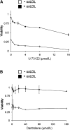
Inhibition of PLC or RyR does not block oxLDL-mediated macrophage survival. A: BMDMs were washed and incubated with media alone or oxLDL (25 µg/ml) in the presence U-73122 or dantrolene at the concentrations indicated for 24 h. B: Viability was measured by the bioreduction of MTS and expressed as a ratio normalized to absorbance values of cells cultured in 10% M-CSF conditioned media.
Fig. 5.
Fig. 5.
S1P generates [Ca2+]i oscillations. Calcium mobilization was visualized using fluo-4-AM and confocal fluorescence microscopy. After incubation with fluo-4-AM, BMDMs were washed, and media containing either nLDL (25 µg/ml), oxLDL (25 µg/ml), or S1P (30 µmol/l) was added at time 0. Fluorescence values as a function of time were measured for every cell in the field, and cells positive for [Ca2+]i oscillations are expressed as a fraction of total cells. ** P < 0.01 compared with cells treated with nLDL.
Fig. 6.
Fig. 6.
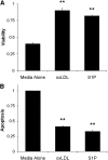
S1P promotes macrophage survival. BMDMs were washed and incubated with media alone, oxLDL (25 µg/ml), or S1P (30 µmol/l) for 24 h. A: Viability was measured by the bioreduction of MTS and expressed as a ratio of normalized to absorbance values of cells cultured in 10% M-CSF conditioned media. B: Apoptosis was assessed by measuring the percentage of cells with subdiploid DNA. Data are expressed as a ratio of normalized to fluorescence values of cells cultured in the absence of M-CSF. ** P < 0.01 compared with cells treated with media alone.
Fig. 7.
Fig. 7.
SK is activated in response to oxLDL. BMDM were washed and incubated in medium without M-CSF for 4 h. OxLDL (25 µg/ml in medium without M-CSF) or oxLDL + SKI (30 µmol/l) was then added for the time periods indicated. SK activity was assessed by measuring the ability of lysates to phosphorylate sphingosine with an in vitro kinase assay. Data are normalized to values of cells at time 0. * P < 0.05 compared with cells at time 0.
Fig. 8.
Fig. 8.
Inhibition of SK blocks oxLDL-generated [Ca2+]i oscillations. Calcium mobilization was visualized using fluo-4-AM and confocal fluorescence microscopy. After incubation with fluo-4-AM, BMDMs were washed, and media containing nLDL (25 µg/ml), oxLDL (25 µg/ml), or oxLDL (25 µg/ml) + SKI (30 µmol/l) was added at time 0. Fluorescence values as a function of time were measured for every cell in the field, and cells positive for [Ca2+]i oscillations are expressed as a fraction of total cells. ** P < 0.01 compared with cells treated with oxLDL alone.
Fig. 9.
Fig. 9.
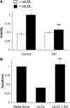
Inhibition of SK blocks oxLDL-mediated macrophage survival. BMDMs were washed and incubated with media alone or oxLDL (25 µg/ml) in the presence or absence of SKI (30 µmol/l) for 24 h. A: Viability was measured by the bioreduction of MTS and expressed as a ratio of normalized to absorbance values of cells cultured in 10% M-CSF conditioned media. B: Apoptosis was assessed by measuring the percentage of cells with subdiploid DNA. Data are expressed as a ratio normalized to fluorescence values of cells cultured in the absence of M-CSF. ** P < 0.01 compared with cells treated with oxLDL alone.

Similar articles

Cited by

References

    1. Ross R. 1999. Atherosclerosis–an inflammatory disease. N. Engl. J. Med. 340: 115–126. - PubMed
    1. Libby P., Theroux P. 2005. Pathophysiology of coronary artery disease. Circulation. 111: 3481–3488. - PubMed
    1. Rosenfeld M. E., Ross R. 1990. Macrophage and smooth muscle cell proliferation in atherosclerotic lesions of WHHL and comparably hypercholesterolemic fat-fed rabbits. Arteriosclerosis. 10: 680–687. - PubMed
    1. Gordon D., Reidy M. A., Benditt E. P., Schwartz S. M. 1990. Cell proliferation in human coronary arteries. Proc. Natl. Acad. Sci. USA. 87: 4600–4604. - PMC - PubMed
    1. Spagnoli L. G., Orlandi A., Santeusanio G. 1991. Foam cells of the rabbit atherosclerotic plaque arrested in metaphase by colchicine show a macrophage phenotype. Atherosclerosis. 88: 87–92. - PubMed

MeSH terms

LinkOut - more resources