. 2010 Jan 28;115(4):860-9.
doi: 10.1182/blood-2009-09-243105. Epub 2009 Nov 25.

Posttranslational stability of the heme biosynthetic enzyme ferrochelatase is dependent on iron availability and intact iron-sulfur cluster assembly machinery

Affiliations

Posttranslational stability of the heme biosynthetic enzyme ferrochelatase is dependent on iron availability and intact iron-sulfur cluster assembly machinery

Daniel R Crooks et al. Blood. .

Abstract

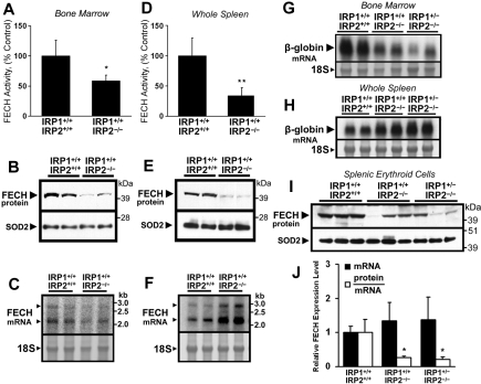
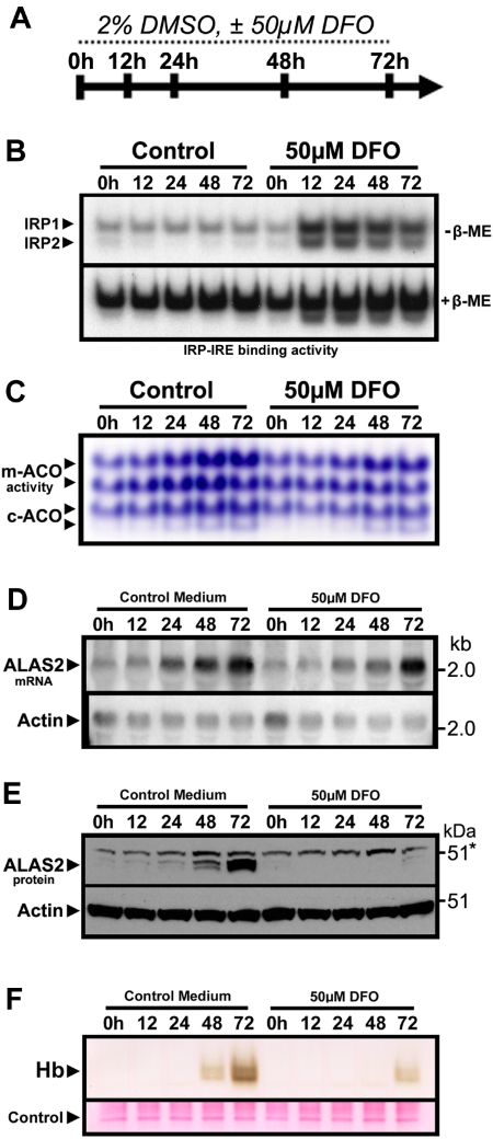
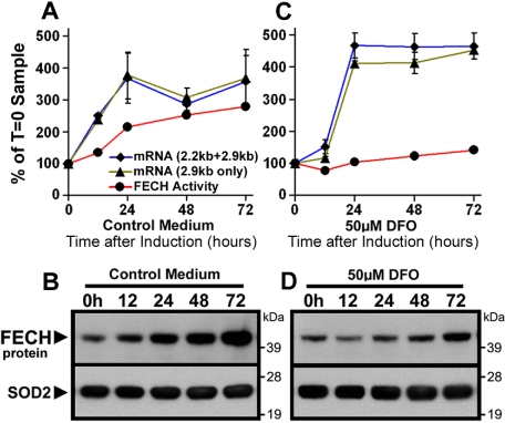
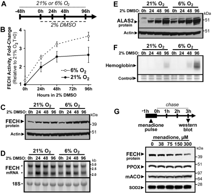
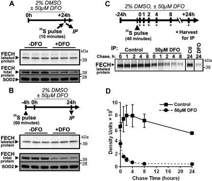
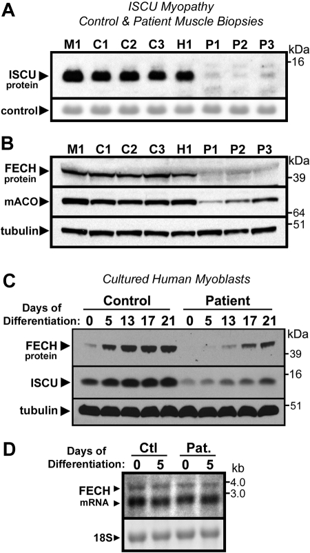
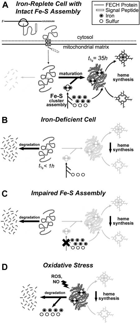
Mammalian ferrochelatase, the terminal enzyme in the heme biosynthetic pathway, possesses an iron-sulfur [2Fe-2S] cluster that does not participate in catalysis. We investigated ferrochelatase expression in iron-deficient erythropoietic tissues of mice lacking iron regulatory protein 2, in iron-deficient murine erythroleukemia cells, and in human patients with ISCU myopathy. Ferrochelatase activity and protein levels were dramatically decreased in Irp2(-/-) spleens, whereas ferrochelatase mRNA levels were increased, demonstrating posttranscriptional regulation of ferrochelatase in vivo. Translation of ferrochelatase mRNA was unchanged in iron-depleted murine erythroleukemia cells, and the stability of mature ferrochelatase protein was also unaffected. However, the stability of newly formed ferrochelatase protein was dramatically decreased during iron deficiency. Ferrochelatase was also severely depleted in muscle biopsies and cultured myoblasts from patients with ISCU myopathy, a disease caused by deficiency of a scaffold protein required for Fe-S cluster assembly. Together, these data suggest that decreased Fe-S cluster availability because of cellular iron depletion or impaired Fe-S cluster assembly causes reduced maturation and stabilization of apo-ferrochelatase, providing a direct link between Fe-S biogenesis and completion of heme biosynthesis. We propose that decreased heme biosynthesis resulting from impaired Fe-S cluster assembly can contribute to the pathogenesis of diseases caused by defective Fe-S cluster biogenesis.


Figures

Figure 1
Figure 1
Posttranscriptional reduction of ferrochelatase (FECH) activity and protein levels in erythropoietic tissues of IRP2−/− mice. (A,D) FECH activity in bone marrow aspirates and whole spleens of Irp1+/+:Irp2−/− mice was significantly decreased compared with Irp1+/+:Irp2+/+ (wild-type) animals. Error bars represent SD (n = 4 animals per genotype). (B,E) FECH protein levels in bone marrow and spleen were also decreased in Irp1+/+:Irp2−/− animals. Each lane from the Western blot represents whole-tissue protein extracts pooled from 2 animals. The filter was reprobed for SOD2 as a loading control for mitochondrial matrix protein. (C,F) FECH mRNA levels were increased in the spleens of Irp1+/+:Irp2−/− mice. Messenger RNA levels were measured by Northern blot using a probe specific for both the 2.2-kb and 2.9-kb FECH transcripts; each lane represents RNA pooled equally from 2 animals. The 18S ribosomal band was visualized to assess equal loading. Results were confirmed by quantitative RT-PCR (data not shown). (G-H) To estimate the tissue distribution of erythroid cells, β-globin mRNA levels in bone marrow aspirates and spleens from Irp1+/+: Irp2+/+, Irp1+/+:Irp2−/−, and Irp1+/−:Irp2−/− mice were assessed by Northern blot. (I) FECH protein abundance in splenic TER119+ erythroid cells mice was measured by Western blot. FECH mRNA transcript levels (J, ■) were measured by quantitative RT-PCR using primers specific for both the 2.2-kb and 2.9-kb FECH transcripts; data are normalized to Irp1+/+:Irp2+/+ (wild-type) mRNA levels. Relative protein abundance was calculated by densitometry, and individual protein abundance values were normalized to the respective mRNA levels measured in the same sample (J, □). Data in panels A, D, and J were analyzed by 2-tailed Student t test; *P < .05; **P < .01.
Figure 2
Figure 2
Iron-limited erythroid differentiation of MEL cells. (A) Timeline depicting the experimental time course of DMSO-induced erythroid differentiation of MEL cells under normal conditions or iron-deficient conditions induced by the iron chelator DFO. (B) Electrophoretic mobility shift assay of MEL cell protein extracts using a 32P-labeled ferritin IRE probe showed activation of IRP1 and IRP2 after treatment with DFO. (C) Aconitase activity gel demonstrating a time-dependent increase in mitochondrial (m-) and cytosolic (c-) aconitase activity levels over the course of differentiation, which was attenuated in DFO-treated cultures. (D) ALAS2 mRNA was induced during differentiation in the presence or absence of DFO. However, ALAS2 protein expression (E) was repressed in DFO-treated cells. Sample loading was assessed by reprobing for actin mRNA and protein, respectively. (F) Mature (heme-containing) hemoglobin (Hb) formation was repressed in differentiating MEL cells treated with DFO. Hemoglobin was measured by a modified diaminobenzidine procedure after separation of total cellular protein (40 μg) by native PAGE followed by transfer to polyvinylidene difluoride filters (“Hemoglobin assay”).
Figure 3
Figure 3
Posttranscriptional reduction of FECH in MEL cells during iron-limited differentiation. (A,C) Induction of FECH enzyme activity (red line), but not mRNA levels (blue, green lines), was repressed during differentiation in the presence of DFO. FECH mRNA levels were measured by quantitative RT-PCR as described in “Northern blots and quantitative RT-PCR analysis.” mRNA transcript and enzyme activity levels are normalized to T = 0 samples. (B,D) Induction of FECH protein levels during differentiation was attenuated in cells cotreated with DFO, despite increased FECH mRNA levels. FECH protein levels were measured by Western blot, and the filter was reprobed for SOD2 as a loading control for mitochondrial matrix proteins.
Figure 4
Figure 4
FECH is destabilized by oxygen and mitochondrial oxidative stress. (A) Cells were maintained in normal (21%) or reduced (6%) O2 atmosphere, followed by differentiation by DMSO treatment. FECH activity (B) and protein levels (C) were increased in differentiating MEL cells maintained at 6% O2 relative to 21% O2 cultures. However, FECH mRNA levels (D) in 6% O2 cultures were not greater than those in 21% O2 cultures. (E) ALAS2 protein expression was also elevated in cells cultured in 6% O2 relative to 21% O2 cultures. (F) Hemoglobinization was accelerated in differentiating cells cultured in 6% O2 (20 μg of total protein was loaded). (G) Menadione-induced mitochondrial oxidative stress caused rapid and specific depletion of total cellular FECH protein levels, whereas PPOX, mACO, and SOD2 protein levels were not appreciably altered. Error bars in panel B represent the range of 2 experimental replicates.
Figure 5
Figure 5
Newly formed, but not mature, FECH protein is susceptible to regulation by iron availability. (A) To test for changes in the instantaneous synthesis rate of FECH protein under iron-depleted conditions, MEL cells were differentiated for 24 hours in the presence or absence of DFO, and harvested immediately after a rapid 10-minute pulse with 35S-Cys and 35S-Met. Radiolabeled FECH was visualized by autoradiography after immunoprecipitation and SDS-PAGE (top panel), whereas total protein levels were measured by Western blot (bottom panel). (B) The effect of iron limitation on mature, Fe-S cluster-containing FECH was assessed by metabolic labeling of cells for 1 hour with 35S-Cys and 35S-Met 4 hours before the onset of 24 hours of differentiation and DFO treatment. After 24-hour incubation, cells were harvested and analyzed for radiolabeled and total protein levels as in panel A. (C) A pulse-chase experiment was performed to follow the fate of newly formed FECH protein during normal and iron-limited growth conditions. After 24-hour differentiation in the presence or absence of DFO, cells were pulsed for 40 minutes with 35S-Cys and 35S-Met, followed by incubation for various periods of time in differentiation medium with or without DFO. A representative autoradiogram is shown; the results of 2 experiments are quantified and plotted in panel D.
Figure 6
Figure 6
Disruption of Fe-S assembly in ISCU myopathy causes depletion of FECH. (A) As reported previously, total ISCU protein levels were depleted in vastus lateralis muscle biopsies taken from Swedish patients with ISCU myopathy (lanes designated as P1-P3), compared with 3 healthy controls (designated C1-C3), and a patient with an unrelated mitochondrial myopathy (designated as M1). A prominent protein band observed after Ponceau-S staining of the filter served as the loading control. (B) FECH protein levels were also greatly decreased in the ISCU myopathy biopsies, as were levels of the Fe-S cluster-containing enzyme mitochondrial aconitase. (C) Primary myotube cultures obtained from an ISCU myopathy patient as well as from a healthy person (control) were terminally differentiated by growth in low serum conditions (“Tissue biopsies and myoblast culture”). Although FECH and ISCU levels increased in both control and patient cultures during the course of the experiment, the relative level of both proteins was diminished in the patient cultures compared with the control at every given time point. (D) Little difference in FECH mRNA levels was seen in control and patient myotube cultures at zero and 5 days of differentiation, as assessed by Northern blot.
Figure 7
Figure 7
The impact of cellular iron deficiency, impaired Fe-S biosynthesis, and mitochondrial oxidative stress on FECH activity and protein levels. (A) A schematic representation of the synthesis, mitochondrial translocation, and maturation of FECH polypeptides under normal conditions. FECH protein is synthesized on cytosolic ribosomes and translocated into the mitochondrion where its signal peptide is cleaved. Complete folding and maturation of FECH requires the provision of a newly formed Fe-S cluster, which is supplied by the Fe-S cluster assembly machinery. After folding and insertion of the cluster, FECH can catalyze the final step in the heme biosynthetic pathway, which is the insertion of ferrous iron into protoporphyrin IX. (B) Under conditions of cellular iron depletion, de novo Fe-S cluster assembly in the mitochondrion is halted because of the lack of available iron ions, and newly imported apo-FECH accumulates and is rapidly degraded within the mitochondrion. (C) Similarly, if mitochondrial Fe-S assembly is disrupted in the absence of cellular iron depletion, as in ISCU myopathy, apo-FECH fails to obtain Fe-S clusters and is rapidly degraded. (D) Under conditions of mitochondrial oxidative stress, mature Fe-S cluster-containing FECH is rapidly destabilized. Degradation is probably initiated by chemical modification or disassembly of the Fe-S cluster, resulting in a conformational change and degradation of the FECH polypeptide.

References

    1. Kikuchi G, Kumar A, Talmage P, Shemin D. The enzymatic synthesis of delta-aminolevulinic acid. J Biol Chem. 1958;233(5):1214–1219. - PubMed
    1. Ajioka RS, Phillips JD, Kushner JP. Biosynthesis of heme in mammals. Biochim Biophys Acta. 2006;1763(7):723–736. - PubMed
    1. Nishida G, Labbe RF. Heme biosynthesis; on the incorporation of iron into protoporphyrin. Biochim Biophys Acta. 1959;31(2):519–524. - PubMed
    1. Cox TC, Bawden MJ, Martin A, May BK. Human erythroid 5-aminolevulinate synthase: promoter analysis and identification of an iron-responsive element in the mRNA. EMBO J. 1991;10(7):1891–1902. - PMC - PubMed
    1. Binder R, Horowitz JA, Basilion JP, Koeller DM, Klausner RD, Harford JB. Evidence that the pathway of transferrin receptor mRNA degradation involves an endonucleolytic cleavage within the 3′-UTR and does not involve poly(A) tail shortening. EMBO J. 1994;13(8):1969–1980. - PMC - PubMed

Publication types

MeSH terms