. 2010 Feb 4;115(5):1088-97.
doi: 10.1182/blood-2009-05-223198. Epub 2009 Dec 4.

Short-term inhibition of p53 combined with keratinocyte growth factor improves thymic epithelial cell recovery and enhances T-cell reconstitution after murine bone marrow transplantation

Affiliations

Short-term inhibition of p53 combined with keratinocyte growth factor improves thymic epithelial cell recovery and enhances T-cell reconstitution after murine bone marrow transplantation

Ryan M Kelly et al. Blood. .

Abstract

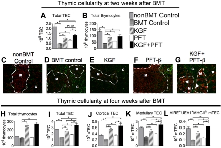
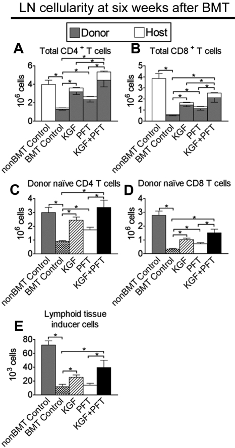
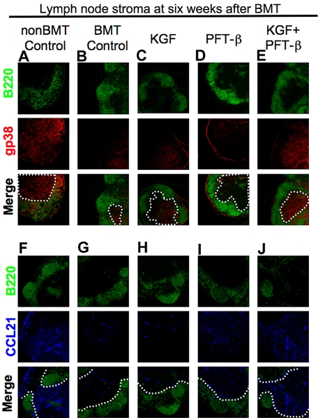
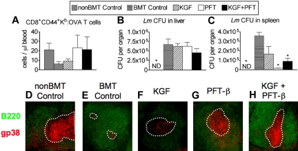
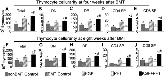
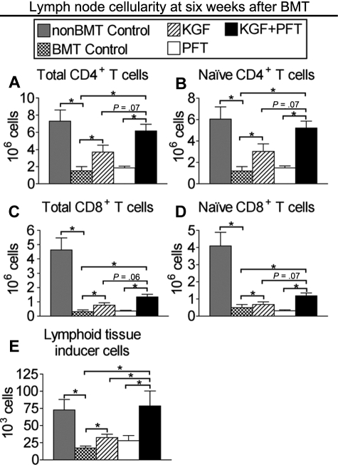
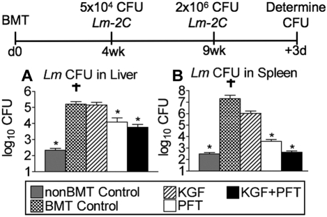
Myeloablative conditioning before bone marrow transplantation (BMT) results in thymic epithelial cell (TEC) injury, T-cell immune deficiency, and susceptibility to opportunistic infections. Conditioning regimen-induced TEC damage directly contributes to slow thymopoietic recovery after BMT. Keratinocyte growth factor (KGF) is a TEC mitogen that stimulates proliferation and, when given before conditioning, reduces TEC injury. Some TEC subsets are refractory to KGF and functional T-cell responses are not fully restored in KGF-treated BM transplant recipients. Therefore, we investigated whether the addition of a pharmacologic inhibitor, PFT-beta, to transiently inhibit p53 during radiotherapy could spare TECs from radiation-induced damage in congenic and allogeneic BMTs. Combined before BMT KGF + PFT-beta administration additively restored numbers of cortical and medullary TECs and improved thymic function after BMT, resulting in higher numbers of donor-derived, naive peripheral CD4(+) and CD8(+) T cells. Radiation conditioning caused a loss of T-cell zone fibroblastic reticular cells (FRCs) and CCL21 expression in lymphoid stroma. KGF + PFT-beta treatment restored both FRC and CCL21 expression, findings that correlated with improved T-cell reconstitution and an enhanced immune response against Listeria monocytogenes infection. Thus, transient p53 inhibition combined with KGF represents a novel and potentially translatable approach to promote rapid and durable thymic and peripheral T-cell recovery after BMT.


Figures

Figure 1
Figure 1
Combined pretreatment with KGF + PFT-β additively restores all TEC subsets by 4 weeks after congenic BMT. Lethally irradiated B6 recipients of congenic (B6 Ly5.1+) BM were left untreated (BMT Control) or pretreated with KGF, PFT-β, or KGF + PFT-β and analyzed for absolute numbers of total (A) TECs (CD45EpCAM+MHC-II+) and (B) thymocytes (SSClowCD45+EpCAMMHC-II) at 2 weeks after BMT. (C-G) Immunofluorescence staining of thymic sections at 2 weeks after BMT for the cytokeratin-5, K5 (red) and Ly51 (green) identified mature cortical TECs (Ly51+K5) and medullary TECs (Ly51K5+). These methods were used to assess recovery of distinct cortical and medullary TEC populations. Images were acquired on an Olympus FV500 confocal microscope with the use of a 10×/0.40 objective lens with associated Olympus Software. (H) Total thymocytes, (I) total TECs, (J) cTECs (CD45 EpCAM+ MHC-II+Ly51+), (K) mTECs (CD45EpCAM+MHC-II+Ly51), and (L) AIRE+ mTEChi (CD45EpCAM+UEA-1highMHC-IIhighLy51 AIRE+) were quantified in thymi from BM transplant recipients and non-BM transplant controls (nonBMT Control) at 4 weeks after BMT. For FACS analysis, data shown are the mean numbers of cells ± SEMs and are representative of 3 experiments of 4 mice per group; *P < .05. For immunofluorescence microscopy, data are representative of 2 experiments, each with 3 mice per group.
Figure 2
Figure 2
Combined pretreatment with KGF + PFT-β additively restores numbers of total and donor-derived, naive CD4+ and CD8+ T cells in LNs by 6 weeks after congenic BMT. Lethally irradiated B6 recipients of congenic (B6 Ly5.1+) BM were left untreated (BMT Control) or pretreated with KGF, PFT-β, or KGF + PFT-β and analyzed for the presence of T cells and Lti cells in the LNs at 6 weeks after BMT alongside unmanipulated age-/sex-matched B6 controls (non-BMT Control). Mean absolute numbers ± SEMs of (A) total CD4+CD3+ T cells; (B) total CD8+CD3+ T cells; (C) donor-derived, naive (CD62LhighCD44low) CD4+CD3+ T cells; (D) donor-derived, naive (CD62LhighCD44low) CD8+CD3+ T cells; and (E) donor-derived, CD4+CD3CD11cB220 Lti cells in the LNs are shown. LN node cells were pooled from inguinal, axillary, and mesenteric LNs. Data are representative of 3 experiments, each with 4 mice per group; *P < .05.
Figure 3
Figure 3
Combined pretreatment with KGF + PFT-β augments T-cell zone FRC and CCL21 expression after congenic BMT. (A-E) Immunofluorescence staining of peripheral LN cryosections for B220+ B cells (green) and gp38+ FRCs (red) was used to assess the relative abundance of gp38+ FRCs in T-cell zones of (A) unmanipulated, age-/sex-matched B6 control (non-BMT Controls) or BM transplant recipients that were (B) left untreated or treated with (C) KGF, (D) PFT-β, or (E) KGF + PFT-β. (F-J) Immunofluorescence staining of LNs for B220 (green) and CCL21 (blue) in (F) unmanipulated, age-/sex-matched B6 control (non-BMT Controls) or BM transplant recipients that were (G) left untreated or treated with (H) KGF, (I) PFT-β, or (J) KGF + PFT-β. In all merged images, a white, dashed line encircles the B220 T-cell zones. Data are representative of 2 experiments, each with 3 mice per group; *P < .05.
Figure 4
Figure 4
Pretreatment with PFT-β or KGF + PFT-β significantly improves primary and secondary immune responses against Lm after congenic BMT. Lethally irradiated B6 recipients of congenic (B6 Ly5.1+) BM were left untreated (BMT Control) or pretreated with KGF, PFT-β, or KGF + PFT-β and immunized at 4 weeks after BMT alongside unmanipulated age-/sex-matched B6 controls (non-BMT control). For primary immunization, 106 CFU of an attenuated strain of Lm that express recombinant full-length chicken ovalbumin (ΔactA-Lm-OVA) was intravenously injected. (A) Absolute numbers of CD44+CD8+ Kb-OVA257-64–specific T cells were quantified in peripheral blood of infected animals by FACS 8 days after primary infection. (B) Immunized mice were then rechallenged with 105 CFU of the virulent parent strain, Lm-OVA, 5 weeks after primary infection. After 3 days, bacterial CFUs in (B) liver and (C) spleen were determined by plating of serial dilutions of organ homogenates onto BHI agar. (D-H) Immunofluorescence staining of spleen cryosections for B220+ B cells (green) and gp38+ FRCs (red) was used to assess the relative abundance of gp38+ FRCs in T-cell zones of (A) unmanipulated, age-/sex-matched B6 control (non-BMT Controls) or BM transplant recipients that were (B) left untreated (BMT control) or treated with (C) KGF, (D) PFT-β, or (E) KGF + PFT-β. A white, dashed line encircles the B220 T-cell zones. Data are representative of 2 experiments, each with 4 mice per group; *P < .05 compared with BMT controls.
Figure 5
Figure 5
Combined pretreatment with KGF + PFT-β additively restores thymocyte cellularity after allogeneic BMT. Lethally irradiated B6 recipients of allogeneic (balb/c) BM were left untreated (BMT control) or pretreated with KGF, PFT-β, or KGF + PFT-β and analyzed for thymocyte cellularity at (A-E) 4 and (F-J) 8 weeks after BMT alongside age-/sex-matched, unmanipulated B6 controls (non-BMT control). Data shown are mean absolute numbers ± SEMs of total thymocytes. The data are pooled from 5 independent experiments with 4 to 5 mice per group; *P < .05 compared with BMT controls; #P < .05 compared with KGF- and PFT-β–treated BM transplant recipients.
Figure 6
Figure 6
Combined pretreatment with KGF + PFT-β additively restores numbers of total and naive CD4+ and CD8+ T cells in LN by 6 weeks after allogeneic BMT. Lethally irradiated CB6F1 recipients of allogeneic (BALB/c) BM were left untreated (BMT Control) or pretreated with KGF, PFT-β, or KGF + PFT-β and analyzed for the presence of T cells in the LNs at 6 weeks after BMT alongside unmanipulated age-/sex-matched CB6F1 controls (non-BMT Control). Mean absolute numbers ± SEMs of (A) total CD4+CD3+ T cells, (B) naive (CD62LhighCD44low) CD4+ T cells, (C) total CD8+CD3+ T cells, (D) naive (CD62LhighCD44low) CD8+ T cells, and (E) CD4+CD3CD11cB220 Lti cells in the LNs are shown. LN cells were pooled from inguinal, axillary, and mesenteric LNs. Data are representative of 2 experiments, each with 4 mice per group; *P < .05.
Figure 7
Figure 7
Pretreatment with KGF and PFT-β significantly improves immune clearance of Lm after allogeneic BMT. Lethally irradiated CB6F1 recipients of allogeneic (balb/c) T cell–depleted BM were left untreated (BMT Control) or pretreated with KGF, PFT-β, or KGF + PFT-β and immunized at 4 weeks after BMT alongside unmanipulated age-/sex-matched CB6F1 controls (non-BMT Control). For primary immunization, 5 × 10⁴ CFU of Lm (strain 2C) was injected intravenously. Immunized mice were rechallenged with 2 × 10⁶ CFU of Lm-2C, 5 weeks after primary infection. Then after 3 days, bacterial CFUs in liver (A) and spleen (B) were determined by plating of serial dilutions of organ homogenates onto BHI agar. Data are representative of 2 experiments, each with 4 mice per group. † indicates mouse succumbed to infection; *P < .05 compared with BMT controls.

Similar articles

Cited by

References

    1. Markova M, Barker JN, Miller JS, et al. Fludarabine vs cladribine plus busulfan and low-dose TBI as reduced intensity conditioning for allogeneic hematopoietic stem cell transplantation: a prospective randomized trial. Bone Marrow Transplant. 2007;39(4):193–199. - PubMed
    1. Weinberg K, Blazar BR, Wagner JE, et al. Factors affecting thymic function after allogeneic hematopoietic stem cell transplantation. Blood. 2001;97(5):1458–1466. - PubMed
    1. Douek DC, Vescio RA, Betts MR, et al. Assessment of thymic output in adults after haematopoietic stem-cell transplantation and prediction of T-cell reconstitution. Lancet. 2000;355(9218):1875–1881. - PubMed
    1. Hakim FT, Memon SA, Cepeda R, et al. Age-dependent incidence, time course, and consequences of thymic renewal in adults. J Clin Invest. 2005;115(4):930–939. - PMC - PubMed
    1. Williams KM, Hakim FT, Gress RE. T cell immune reconstitution following lymphodepletion. Semin Immunol. 2007;19(5):318–330. - PMC - PubMed

Publication types

MeSH terms