. 2010 Mar 25;115(12):2380-90.
doi: 10.1182/blood-2009-07-233031. Epub 2009 Nov 13.

gp96, an endoplasmic reticulum master chaperone for integrins and Toll-like receptors, selectively regulates early T and B lymphopoiesis

Affiliations

gp96, an endoplasmic reticulum master chaperone for integrins and Toll-like receptors, selectively regulates early T and B lymphopoiesis

Matthew Staron et al. Blood. .

Abstract

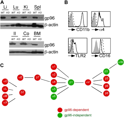
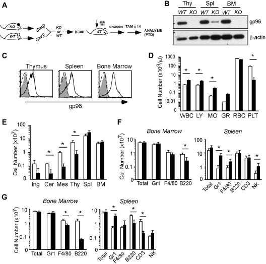
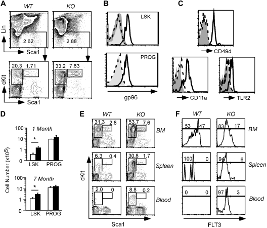
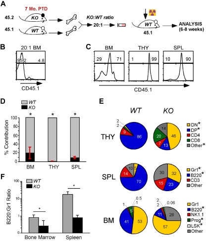
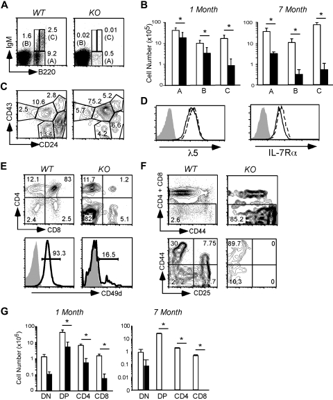
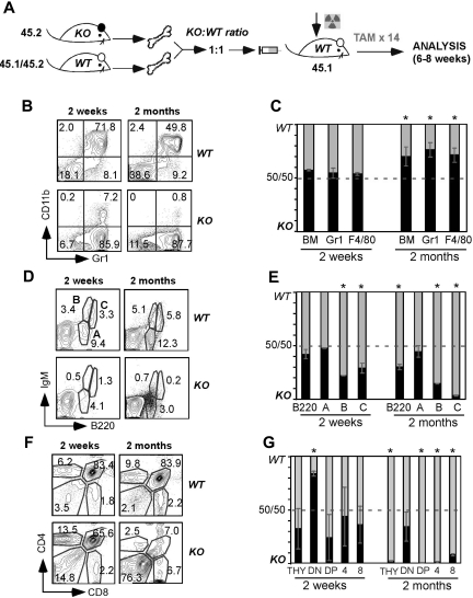
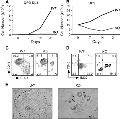
Integrins contribute to lymphopoiesis, whereas Toll-like receptors (TLRs) facilitate the myeloid replenishment during inflammation. The combined role of TLRs and integrin on hematopoiesis remains unclear. gp96 (grp94, HSP90b1) is an endoplasmic reticulum master chaperone for multiple TLRs. We report herein that gp96 is also essential for expression of 14 hematopoietic system-specific integrins. Genetic deletion of gp96 thus enables us to determine the collective roles of gp96, integrins, and TLRs in hematopoiesis. We found that gp96-null hematopoietic stem cells could support long-term myelopoiesis. B- and T-cell development, however, was severely compromised with transitional block from pro-B to pre-B cells and the inability of thymocytes to develop beyond the CD4(-)CD8(-) stage. These defects were cell-intrinsic and could be recapitulated on bone marrow stromal cell culture. Furthermore, defective lymphopoiesis correlated strongly with failure of hematopoietic progenitors to form close contact with stromal cell niche and was not the result of the defect in the assembly of antigen receptor or interleukin-7 signaling. These findings define gp96 as the only known molecular chaperone to specifically regulate T- and B-cell development.


Figures

Figure 1
Figure 1
Inducible deletion of gp96 reveals its critical role for expression of multiple integrins. WT or gp96 KO mice were injected with tamoxifen (TAM) intraperitoneally for 14 days followed by analysis. (A) Immunoblot for gp96 and β-actin (a loading control) from multiple organs: Li indicates liver; Lu, lung; Ki, kidney; Spl, spleen; Il, ileum; and Co, colon. (B) Flow cytometric analysis of BM Gr1+ cells for cell-surface expression of indicated molecules. Dotted histogram represents KO cells; solid line indicates WT cells. (C) Summary of gp96-dependent and independent integrins on the hematopoietic system. These data are based on numerous experiments.
Figure 2
Figure 2
Global consequence of gp96 deletion on hematopoiesis. (A) Experimental scheme. (B) Immunoblot of various hematopoietic tissues from WT and KO mice 12 days post-TAM deletion (PTD). Thy indicates thymus. β-Actin serves as loading control. (C) Intracellular staining for gp96 in various hematopoietic tissues from WT (solid line) or KO (dotted line) bone marrow (BM) chimeras using gp96 antibody (open) or isotype control (shaded). (D) Peripheral blood cell counts of WT (n = 3, □) and KO (n = 4, ■) BM chimeras 1 month PTD. (E) Organ cellularity of WT (n = 3, □) and KO (n = 4, ■) BM chimeras 1 month PTD. Ing indicates inguinal; Cer, cervical; and Mes, mesenteric. (F-G) Absolute cell numbers of indicated lineages in BM and spleen of WT (n = 5, □) and KO (n = 6, ■) BM chimeras 3 to 4 weeks PTD (F) and 7 months PTD (G). Data are representative of at least 3 independent experiments. WBC indicates white blood cell; LY, lymphocyte; MO, monocyte; GR, granulocyte; RBC, red blood cell; and PLT, platelet. *P < .05.
Figure 3
Figure 3
Multipotent hematopoietic progenitor persists in the absence of gp96. (A top) Dump gating of BM cells from WT and KO BM chimeras 1 month PTD. (Bottom) Gating of LSK (L) and PROG (P) populations. Numbers represent percentage of cells in the gated population. (B) Intracellular staining of gp96 on BM LSK and PROG populations from WT (solid line) and KO (dotted line) BM chimeras 2 months PTD using polyclonal gp96 antibody or isotype control (shaded). (C) CD49d, CD11a, and TLR2 expression on BM LSK populations from WT (solid line) and KO (dotted line) BM chimeras 1 month PTD. (D) Absolute numbers of BM LSK and PROG from WT (n = 3, □) and KO (n = 4, ■) BM chimeras 1 month and 7 months PTD. *P < .05. (E) LSK and PROG analysis from WT and KO BM chimeras 2 months PTD in the indicated tissues. (F) FLT3 expression on LSK from WT and KO BM chimeras 2 months PTD in the indicated tissues. Data are representative of at least 2 independent experiments.
Figure 4
Figure 4
De novo lymphopoiesis, but not myelopoiesis, is dependent on gp96. (A) Diagram of experimental design: BM cells from KO (7 months PTD BM chimera, 45.2) and WT (45.1) mice were transplanted in 20:1 ratio to lethally irradiated recipients (45.1) and analyzed 4 to 6 weeks PTD. (B) Representative congenic analysis of donor BM after 20:1 mix of WT and KO cells. (C) Congenic analysis of a representative recipient for relative contribution by WT (n = 4) and KO (n = 4) cells to the various hematopoietic tissues 4 to 6 weeks after BM reconstitution. (D-E) Congenic gated WT (gray) and KO (black) cells were analyzed for their relative contribution (number indicates percentage) to various lineages and hematopoietic tissues. (F) Percentage of B220+ cells over Gr1+ cells in BM and spleen of WT and KO mice. Data are representative of 2 independent experiments. *P < .05.
Figure 5
Figure 5
B- and T-cell lymphopoiesis is dependent on gp96 at critical transitional stages. (A) B220/IgM surface staining of BM cells from WT and KO BM chimeras 1 month PTD. (B) Absolute numbers of BM B cells in various fractions indicated in panel A from WT (n = 5, □) and KO (n = 6, ■) BM chimeras 3 to 4 weeks PTD. Data are pooled from 2 independent experiments. (C) CD43/CD24 surface staining on B220+ cells in BM. (D) Expression of λ5 pre-BCR and IL-7Rα on the surface of WT (solid line) and KO (dotted line) pre–B-cell line. Shaded histograms represent isotype control. (E) CD4/CD8 (top) surface staining and surface expression of CD49d (bottom) on thymocytes from WT and KO BM chimeras 1 month PTD. (F) DN gating (top) and CD44/CD25 surface staining on DN thymocytes from WT and KO BM chimeras 1 month PTD. (G) Absolute cell numbers of thymocytes from the indicated stages from WT (n = 3, □) and KO (n = 4, ■) BM chimeras 1 month and 7 months PTD. Data are representative of at least 2 independent experiments. *P < .05.
Figure 6
Figure 6
Competitive BM reconstitution reveals cell-intrinsic role for gp96 in B and T lymphopoiesis, but not myelopoiesis. (A) Diagram of experimental design. (B,D,F) Surface staining of the indicated lineages on gated KO (45.2) and WT (45.1/45.2) BM cells from 1:1 BM chimeras 2 weeks and 2 months PTD. (C,E,G) Percentage of KO and WT contribution to total BM myeloid cells (C), BM B-cell subsets (E), and thymocytes (G) at 2 weeks and 2 months PTD (DN: CD4CD8; double-positive: CD4+CD8+; 4: CD4+CD8; 8: CD4CD8+). Data are representative of 3 mice per group. *P < .05.
Figure 7
Figure 7
gp96 null hematopoietic progenitors fail to proliferate and differentiate in BM stromal cell cultures. Purified WT and gp96 KO BM Linc-kit+ progenitor cells were cultured on BM stromal cell OP9 or OP9-DL1 for 3 weeks and analyzed for cell growth and differentiation. (A-B) Kinetic analysis of cell proliferation. (C-D) Flow cytometric analysis of T- and B-cell development after 3 weeks of coculture. (E) Bright field image of day 5 coculture of WT or KO Linc-kit+ progenitors on OP9 (original magnification ×100). Dotted circle denotes “cobblestone” colony. A few KO progenitors were indicated by. Three experiments were performed with similar results.

Comment in

References

    1. Miranti CK, Brugge JS. Sensing the environment: a historical perspective on integrin signal transduction. Nat Cell Biol. 2002;4(4):E83–E90. - PubMed
    1. Luo BH, Carman CV, Springer TA. Structural basis of integrin regulation and signaling. Annu Rev Immunol. 2007;25:619–647. - PMC - PubMed
    1. Arroyo AG, Yang JT, Rayburn H, Hynes RO. Differential requirements for alpha4 integrins during fetal and adult hematopoiesis. Cell. 1996;85(7):997–1008. - PubMed
    1. Arroyo AG, Yang JT, Rayburn H, Hynes RO. Alpha4 integrins regulate the proliferation/differentiation balance of multilineage hematopoietic progenitors in vivo. Immunity. 1999;11(5):555–566. - PubMed
    1. Scott LM, Priestley GV, Papayannopoulou T. Deletion of alpha4 integrins from adult hematopoietic cells reveals roles in homeostasis, regeneration, and homing. Mol Cell Biol. 2003;23(24):9349–9360. - PMC - PubMed

Publication types

MeSH terms