. 2010 Jan 21;115(3):559-69.
doi: 10.1182/blood-2009-07-235747. Epub 2009 Nov 19.

Dual targeting of the PI3K/Akt/mTOR pathway as an antitumor strategy in Waldenstrom macroglobulinemia

Affiliations

Dual targeting of the PI3K/Akt/mTOR pathway as an antitumor strategy in Waldenstrom macroglobulinemia

Aldo M Roccaro et al. Blood. .

Abstract

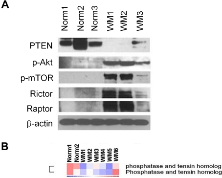
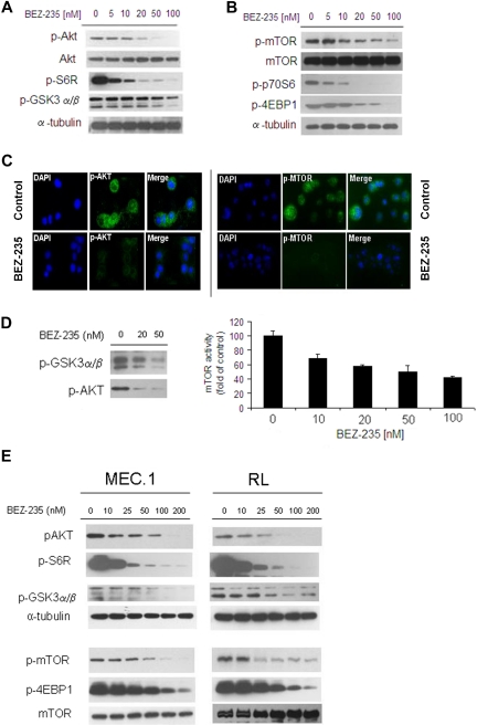
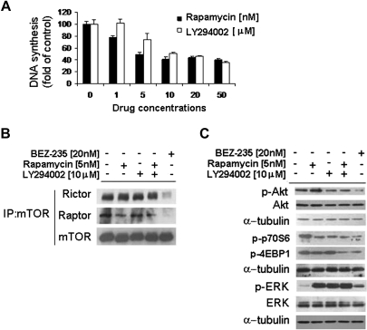
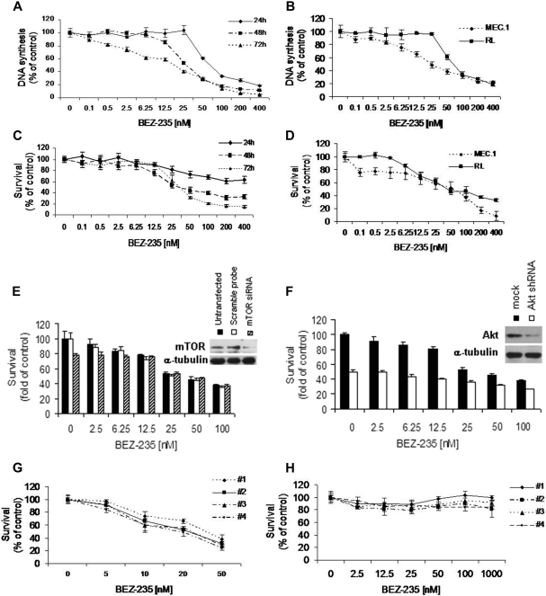
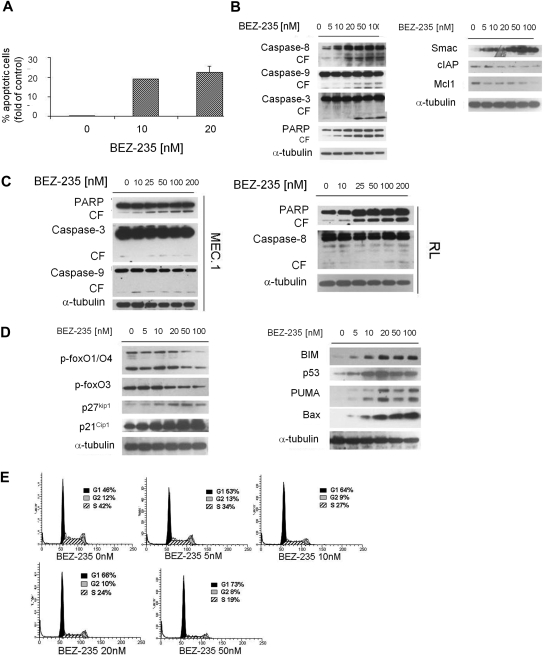
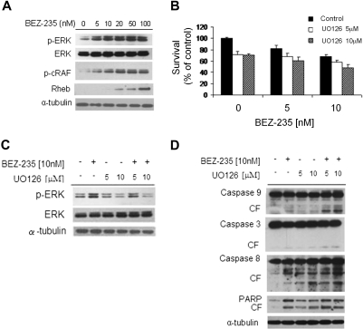
We have previously shown clinical activity of a mammalian target of rapamycin (mTOR) complex 1 inhibitor in Waldenstrom macroglobulinemia (WM). However, 50% of patients did not respond to therapy. We therefore examined mechanisms of activation of the phosphoinositide 3-kinase (PI3K)/Akt/mTOR in WM, and mechanisms of overcoming resistance to therapy. We first demonstrated that primary WM cells show constitutive activation of the PI3K/Akt pathway, supported by decreased expression of phosphate and tensin homolog tumor suppressor gene (PTEN) at the gene and protein levels, together with constitutive activation of Akt and mTOR. We illustrated that dual targeting of the PI3K/mTOR pathway by the novel inhibitor NVP-BEZ235 showed higher cytotoxicity on WM cells compared with inhibition of the PI3K or mTOR pathways alone. In addition, NVP-BEZ235 inhibited both rictor and raptor, thus abrogating the rictor-induced Akt phosphorylation. NVP-BEZ235 also induced significant cytotoxicity in WM cells in a caspase-dependent and -independent manner, through targeting the Forkhead box transcription factors. In addition, NVP-BEZ235 targeted WM cells in the context of bone marrow microenvironment, leading to significant inhibition of migration, adhesion in vitro, and homing in vivo. These studies therefore show that dual targeting of the PI3K/mTOR pathway is a better modality of targeted therapy for tumors that harbor activation of the PI3K/mTOR signaling cascade, such as WM.


Figures

Figure 1
Figure 1
Primary WM cells are characterized by low expression of PTEN and high expression of p-Akt and p-mTOR, compared with their normal cellular counterpart. (A) Immunoblotting for PTEN, p-Akt, p-mTOR, rictor, raptor, and β-actin expression in BM-derived CD19+ WM primary cells (WM), compared with normal primary CD19+ cells (Norm). (B) Purified cRNA (15 μg) isolated from primary BM-derived CD19+ WM primary cells (WM) and normal primary CD19+ cells (Norm) was hybridized to HG-U133Plus2.0 GeneChip (Affimetrix). Fold change is shown by the intensity of induction (red) or suppression (blue).
Figure 2
Figure 2
NVP-BEZ235 targets Akt and mTOR, and inhibits Akt and mTOR activity in WM cells and other low-grade lymphoma IgM-secreting cell lines. (A-B) BCWM.1 cells were cultured with NVP-BEZ235 (5-100nM) for 6 hours. Whole-cell lysates were subjected to Western blotting using anti–p-Akt, anti-Akt, anti–p-S6R, anti–p-GSK3α/β, anti–p-mTOR, anti-mTOR, p-p70S6, anti–p-4EBP1, and anti–α-tubulin antibodies. (C) BCWM.1 cells were cultured with NVP-BEZ235 (20nM) or control medium for 6 hours. Immunocytochemical analysis was assessed using anti–p-Akt and p-mTOR antibodies. The 4′,6-diamidino-2-phenylindole was used to stain nuclei. (D) In vitro Akt and in vitro mTOR kinase assays. BCWM.1 cells were cultured with control media or NVP-BEZ235 (20-50nM) for 6 hours. Akt kinase assay: whole-cell lysates were immunoprecipitated with anti-Akt antibody. Then the immunoprecipitate was washed and subjected to in vitro kinase assay, according to the manufacturer's protocol. Western blotting used anti–p-GSK3α/β and anti-Akt antibodies. mTOR kinase assay: whole-cell lysates were immunoprecipitated with anti-mTOR antibody. Then the immunoprecipitate was washed and subjected to in vitro kinase assay, according to the manufacturer's protocol. Enzyme-linked immunosorbent assay–based assay has been performed (anti-p70S6K was added to each well), and absorbance read at 450nM with a reference wavelength set at 540nM. (E) IgM-secreting cell lines (MEC.1; RL) were cultured with NVP-BEZ235 (10-200nM) for 6 hours. Whole-cell lysates were subjected to Western blotting using anti–p-Akt, anti-Akt, anti–p-S6R, anti–p-GSK3α/β, anti–p-mTOR, anti–p-4EBP1, anti-mTOR, and anti–α-tubulin antibodies.
Figure 3
Figure 3
NVP-BEZ235 targets both rictor and raptor. (A) Thymidine uptake assay. BCWM.1 cells were cultured with rapamycin and LY294002 for 48 hours. (B-C) BCWM.1 cells were cultured with NVP-BEZ235 (20nM), rapamycin (5nM), LY294002 (10μM), or the combination of rapamycin and LY294002 for 6 hours. (B) Whole-cell lysates were subjected to immunoprecipitation (IP) using a mTOR antibody and Western blotting using anti-mTOR, anti-rictor, and anti-raptor antibodies. (C) Whole-cell lysates were subjected to Western blot using anti–p-Akt, anti-Akt, anti–p-p70S6, anti–p-4EBP1, anti–p-ERK, anti-ERK, and anti–α-tubulin antibodies.
Figure 4
Figure 4
NVP-BEZ235 decreases DNA synthesis and triggers cytotoxicity in WM cells and IgM-secreting cell lines, without affecting normal cells. (A) Thymidine uptake assay. BCWM.1 cells were cultured with NVP-BEZ235 (0.1-400nM) for 24 to 72 hours. (B) Thymidine uptake assay. IgM-secreting cell lines (MEC.1; RL) were cultured with NVP-BEZ235 (0.1-400nM) for 48 hours. (C) Cytotoxicity was assessed by MTT assay. BCWM.1 cells were cultured with BEZ235 (0.1-400nM) for 24 to 48 hours and 48 to 72 hours. (D) Cytotoxicity was assessed by MTT assay. IgM-secreting cell lines (MEC.1; RL) were cultured with NVP-BEZ235 (0.1-400nM) for 48 hours. (E) BCWM.1 cells were transfected with either scramble probe or mTOR siRNA. Untransfected BCWM.1 cells were used as control. Cells were treated with NVP-BEZ235 (2.5-100nM) for 48 hours, and cytotoxicity was assessed by MTT assay. Whole-cell lysates were subjected to Western blotting using anti-mTOR and α-tubulin antibodies (inset [E]). (F) BCWM.1 cells were transduced with Akt short hairpin RNA. Mock: control plasmid. BCWM.1-transfected cells or BCWM.1 control cells were treated with NVP-BEZ235 (2.5-100nM) for 48 hours, and cytotoxicity was assessed by MTT. Whole-cell lysates were subjected to Western blotting using anti-Akt and α-tubulin antibodies (inset [F]). (G) Freshly isolated BM CD19+ tumor cells from 4 patients with WM were cultured with NVP-BEZ235 (5-50nM) for 48 hours. Cytotoxicity was assessed by MTT assay. (H) Freshly isolated peripheral blood–derived CD19+ from 4 healthy donors was cultured with NVP-BEZ235 (2.5-1000nM) for 48 hours.
Figure 5
Figure 5
NVP-BEZ235 induces apoptosis and cell-cycle arrest in WM cells. (A) BCWM.1 cells were cultured with NVP-BEZ235 (10-20nM) for 48 hours, and apoptosis was performed using annexin/PI staining and flow cytometric analysis. (B) BCWM.1 cells were cultured with NVP-BEZ235 (5-100nM) for 16 hours. Whole-cell lysates were subjected to Western blotting using anti–caspase-8, anti–caspase-9, anti–caspase-3, anti-PARP, anti-Smac/DIABLO, anti–c-IAP1, anti-Mcl1, and anti–α-tubulin antibodies. (C) IgM-secreting cell lines (MEC.1; RL) were cultured with NVP-BEZ235 (5-100nM) for 16 hours. Whole-cell lysates were subjected to Western blotting using anti–caspase-8, anti–caspase-9, anti–caspase-3, anti-PARP, and anti–α-tubulin antibodies. (D) BCWM.1 cells were cultured with NVP-BEZ235 (5-100nM) for 16 hours. Whole-cell lysates were subjected to Western blotting using anti–p-foxO1/O4, anti–p-foxO3, anti-p27kip1, anti-p21Cip1, anti-BIM, anti-p53, anti-PUMA, anti-Bax, and anti–α-tubulin antibodies. (E) BCWM.1 were cultured with NVP-BEZ235 (0-50nM) for 24 hours, and cell-cycle analysis was performed by PI staining.
Figure 6
Figure 6
NVP-BEZ235 up-regulates the MEK/ERK pathway, and ERK inihibition increases NVP-BEZ235–induced cytotoxicity. (A) BCWM.1 cells were cultured with NVP-BEZ235 (5-100nM) for 6 hours. Whole-cell lysates were subjected to Western blotting using anti–p-ERK, anti-ERK1/2, anti–p-c-Raf, anti-Rheb, and α-tubulin antibodies. (B) BCWM.1 cells were cultured for 48 hours with NVP-BEZ235 (5nM, 10nM) in the absence or presence of MEK1/2 inhibitor U0126 (5μM, 10μM). Cytotoxicity was assessed by the MTT assay. Data represent mean (± SD) of triplicate experiments. (C) BCWM.1 cells were cultured with NVP-BEZ235 (5nM, 10nM) in the absence or presence of MEK1/2 inhibitor U0126 (5μM, 10μM) for 6 hours. Whole-cell lysates were subjected to Western blotting using anti–p-ERK, anti-ERK1/2, and α-tubulin antibodies. (D) BCWM.1 cells were cultured with NVP-BEZ235 (5nM, 10nM) in the absence or presence of MEK1/2 inhibitor U0126 (5μM, 10μM) for 16 hours. Whole-cell lysates were subjected to Western blotting using anti–caspase-9, anti–caspase-3, anti–caspase-8, anti-PARP, and anti–α-tubulin antibodies.
Figure 7
Figure 7
Coculture with BMSCs does not protect against NVP-BEZ235–induced WM cell cytotoxicity. (A) BCWM.1 cells were cultured with NVP-BEZ235 (6.25-100nM) for 48 hours in the presence or absence of BMSCs. Cell proliferation was assessed using the [3H]-thymidine uptake assay. (B) BCWM.1 cells were cultured with either NVP-BEZ235 (5-100nM) alone or in presence of BMSCs for 6 hours. Whole-cell lysates were subjected to Western blotting using anti–p-Akt, anti-Akt, p-mTOR, anti-raptor, anti-rictor, and anti–α-tubulin antibodies. (C) Adhesion assay with BCWM.1 in the presence or absence of NVP-BEZ235 (5-50nM). BCWM.1 demonstrated increased adhesion in FN-coated wells compared with control. NVP-BEZ235 inhibited adhesion in FN-coated wells in a dose-dependent manner (P < .05). All data represent mean (± SD) of triplicate experiments. Adhesion of BCWM.1 cell to primary BMSCs. BCWM.1 demonstrated increased adhesion in BMSC-coated wells compared with control. NVP-BEZ235 inhibited adhesion to BMSCs in a dose-dependent manner (P < .02). All data represent mean (± SD) of triplicate experiments. (D) Transwell migration assay. SDF-1 (30nM) was placed in the lower chambers, and migration was determined after 3 hours. Transwell migration assay showing inhibition of migration of BCWM.1 in the presence of NVP-BEZ235 (5-50nM). SDF-1 (30nM) was placed in the lower chambers and induced migration compared with control with no SDF-1. NVP-BEZ235 inhibited SDF-1–induced migration in WM cells (P < .05). (E) BCWM.1 cells were cultured with NVP-BEZ235 (6.25-100nM) for 4 hours. Whole-cell lysates were subjected to Western blotting using anti–p-focal adhesion kinase, anti–p-paxillin, anti–p-cofilin, and anti–α-tubulin antibodies. (F) In vivo flow cytometry. Calcein red/orange–labeled cells treated with 20nM NVP-BEZ235 and calcein green/AM–labeled, untreated cells were injected in the tail vein of 2 BALB/c mice. Cells were counted every 5 minutes for 60 minutes. Fluorescence signal was detected on an appropriate artery in the ear and digitized for analysis with Matlab software (*P < .05).

Similar articles

Cited by

References

    1. Vivanco I, Sawyers CL. The phosphatidylinositol 3-kinase AKT pathway in human cancer. Nat Rev Cancer. 2002;2:489–501. - PubMed
    1. Pene F, Claessens YE, Muller O, et al. Role of the phosphatidylinositol 3-kinase/Akt and mTOR/P70S6-kinase pathways in the proliferation and apoptosis in multiple myeloma. Oncogene. 2002;21:6587–6597. - PubMed
    1. Leleu X, Jia X, Runnel J, et al. The Akt pathway regulates survival and homing in Waldenstrom macroglobulinemia. Blood. 2007;110:4417–4426. - PMC - PubMed
    1. Neshat MS, Mellinghoff IK, Tran C, et al. Enhanced sensitivity of PTEN-deficient tumors to inhibition of FRAP/mTOR. Proc Natl Acad Sci U S A. 2001;98:10314–10319. - PMC - PubMed
    1. Van de Sande T, De Schrijver E, Heyns W, Verhoeven G, Swinnen JV. Role of the phosphatidylinositol 3′-kinase/PTEN/Akt kinase pathway in the overexpression of fatty acid synthase in LNCaP prostate cancer cells. Cancer Res. 2002;62:642–646. - PubMed

Publication types

MeSH terms