. 2010 Mar 25;115(12):2520-32.
doi: 10.1182/blood-2009-07-233478. Epub 2009 Nov 23.

Vascular endothelial growth factor activation of endothelial cells is mediated by early growth response-3

Affiliations

Vascular endothelial growth factor activation of endothelial cells is mediated by early growth response-3

Jun-ichi Suehiro et al. Blood. .

Abstract

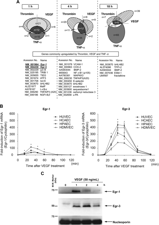
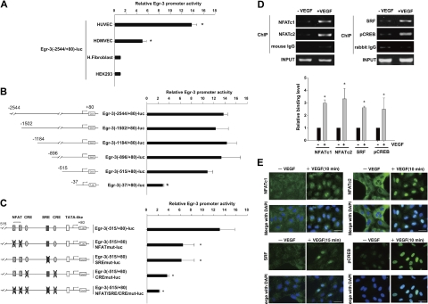
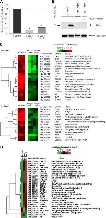
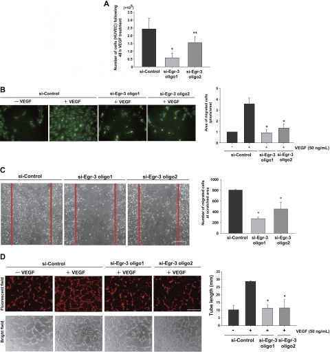
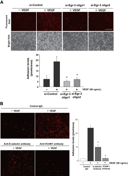
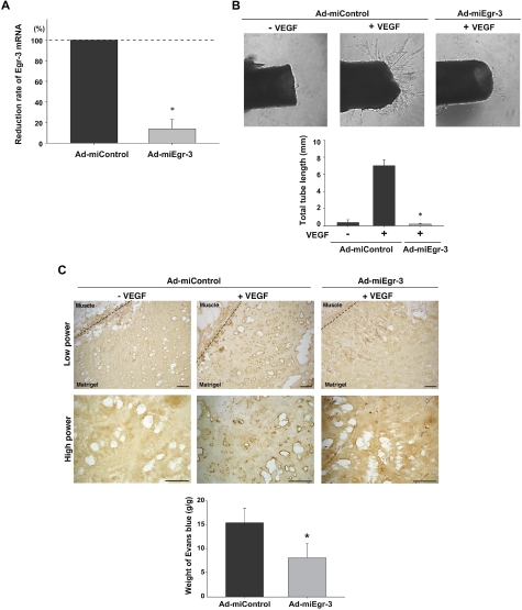
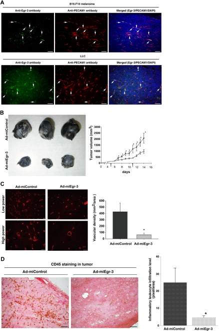
Endothelial cell activation and dysfunction underlie many vascular disorders, including atherosclerosis, tumor growth, and sepsis. Endothelial cell activation, in turn, is mediated primarily at the level of gene transcription. Here, we show that in response to several activation agonists, including vascular endothelial growth factor (VEGF), tumor necrosis factor-alpha, and thrombin, endothelial cells demonstrate rapid and profound induction of the early growth response (Egr) genes egr-1 and egr-3. In VEGF-treated endothelial cells, induction of Egr-3 was far greater and more prolonged compared with Egr-1. VEGF-mediated stimulation of Egr-3 involved the inducible binding of NFATc, serum response factor, and CREB to their respective consensus motifs in the upstream promoter region of Egr-3. Knockdown of Egr-3 markedly impaired VEGF-mediated proliferation, migration, and tube formation of endothelial cells and blocked VEGF-induced monocyte adhesion. Egr-3 knockdown abrogated VEGF-mediated vascular outgrowth from ex vivo aortic rings and attenuated Matrigel plug vascularization and melanoma tumor growth in vivo. Together, these findings suggest that Egr-3 is a critical determinant of VEGF signaling in activated endothelial cells. Thus, Egr-3 represents a potential therapeutic target in VEGF-mediated vasculopathic diseases.


Figures

Figure 1
Figure 1
Activation agonists result in rapid, high-level induction of Egr-3 in primary human endothelial cells. (A) DNA microarrays of HUVECs treated with 50 ng/mL VEGF, 2 U/mL thrombin, or 10 ng/mL TNF-α for 1, 4 or 18 hours. Shown is Venn diagram of agonist-induced genes. (B) Real-time PCR time-course analysis of Egr-1 and -3 mRNA expression in VEGF-treated HUVECs, HCAECs, HPAECs, and HDMVECs. *P < .001 compared with 0 hours (no treatment) in HUVECs, HCAECs, HPAECs, and HDMVECs. (C) Time-dependent induction of Egr-1 and -3 protein expression in VEGF-stimulated HUVECs. Western blot analysis was performed by the use of antibodies against Egr-1 or -3. Antinucleoporin antibody was used as an internal loading control. The data are representative of 3 independent experiments.
Figure 2
Figure 2
NFAT, SRF, and CREB bind to and transactivate the Egr-3 promoter in primary human endothelial cells. (A) Egr-3 (−2544/+80)-luc was transiently transfected into HUVECs, HDMVECs, human skin fibroblasts (H.Fibroblast), or HEK-293 cells. Cells were treated in the absence or presence of 50 ng/mL VEGF for 4 hours and assayed for reporter gene activity. The results show the mean ± SD of luciferase light units in VEGF-treated cells relative to untreated cells, obtained in triplicate from 3 independent experiments. *P < .001 compared with untreated cells. (B) 5′-deletion analysis of Egr-3 promoter activity in control versus VEGF-treated HUVECs. Successive deletions of the 5′-flanking region of Egr-3 were coupled to luciferase in pGL3, and the resulting constructs were transiently transfected into HUVECs. Cells were treated in the absence or presence of 50 ng/mL VEGF for 4 hours and assayed for reporter gene activity. The results show the mean ± SD of luciferase light units in VEGF-treated cells relative to untreated cells obtained in triplicate from 3 independent experiments. *P < .01 compared with Egr-3 (−2544/+80)-luc. (C) Analysis of mutant Egr-3 promoter activity in control versus VEGF-treated HUVECs. HUVECs were transiently transfected with either wild-type Egr-3 (−515/+80)-luc or Egr-3 (−515/+80)-luc containing point mutations of NFAT, SRF, and/or CRE motifs; treated in the absence or presence of 50 ng/mL VEGF for 4 hours; and assayed for reporter gene activity. The results show the mean ± SD of luciferase light units in VEGF-treated cells relative to untreated cells obtained in triplicate from 3 independent experiments. *P < .01 compared with Egr-3 (−515/+80)-luc. (D) ChIP assays of HUVECs treated in the absence or presence of 50 ng/mL VEGF for 30 minutes. Formalin-fixed chromatin was immunoprecipitated with monoclonal mouse antibodies to NFATc1 or NFATc2 or with mouse control immunoglobulin (IgG; left). Alternatively, formalin-fixed chromatin was immunoprecipitated with rabbit polyclonal antibodies against SRF or phospho-CREB or with rabbit control IgG (right). Precipitated chromatin was PCR amplified (30-35 cycles) and subjected to agarose gel electrophoresis (top). Binding was quantified by the use of real-time PCR (bottom). The results show the mean ± SE of binding level relative to control (without VEGF) obtained from at least 3 independent experiments. *P < .01 compared with control. (E) Immunofluorescent studies of NFAT, SRF, and phospho-CREB (pCREB) in control and VEGF-treated HUVECs. Serum-starved cells were incubated in the presence or absence of 50 ng/mL VEGF for the times indicated. The cells were then fixed and incubated with antibodies against NFATc1, NFATc2, SRF, or phospho-CREB, followed by Alexa 488–conjugated second antibody (green). The nuclei were stained with DAPI (blue). Merged images are shown in the bottom row. White bar indicates 20 μm.
Figure 3
Figure 3
Egr-3 is required for VEGF-mediated induction of proangiogenic and proinflammatory genes in primary human endothelial cells. (A) Quantitative real-time PCR analysis of Egr-3 mRNA expression in HUVECs transfected with control siRNA (si-Control) or 2 independent siRNAs against Egr-3 (si-Egr-3 oligo1 and -2) and then treated in the presence of 50 ng/mL VEGF for 1 hour. Egr-3 expression is normalized to Cyclophilin A mRNA levels. The results show the mean ± SD of expression relatives relative to si-Control obtained from at least 5 independent experiments. *P < .001 compared with si-Control. (B) Western blot analysis of Egr-3 protein in HUVECs transfected with control siRNA or 2 independent siRNAs against Egr-3 and treated in the absence of presence of 50 ng/mL VEGF for 1 hour. Nucleoporin was used as a loading control. The data are representative of 4 independent experiments. (C) DNA microarrays of HUVECs transfected with si-Control, si-Egr-3 oligo1, or si-Egr-3 oligo2 and treated with 50 ng/mL VEGF for 1 hour. Shown are heat maps of genes whose VEGF response was most profoundly inhibited by si-Egr-3. Transcriptome data were derived from triplicate arrays of VEGF-treated si-Control–transfected cells and duplicate arrays of each of the VEGF-treated si-Egr-3–transfected cells. (D) DNA microarrays of HUVECs transduced with Ad-control or Ad-expressing Egr-1 (Ad-Egr-1) or Egr-3 (Ad-Egr-3; multiplicity of infection = 5). Cells were harvested for RNA at 24 hours after infection. Shown is heat map of siEgr-3–mediated down-regulated genes in Ad-Egr vs Ad-control–transduced cells.
Figure 4
Figure 4
Egr-3 is required for VEGF-mediated induction of proliferation, migration, and tube formation of primary human endothelial cells. (A) Proliferation assay. HUVECs (105) were transfected with si-Control, si-Egr-3 oligo1, or si-Egr-3 oligo2. Cells were serum-starved for 18 hours, incubated in the presence of 50 ng/mL VEGF for 48 hours, and subsequently enumerated. The results show the mean ± SD derived from 6 independent experiments. *P < .01; **P < .04 compared with si-Control. (B) Migration assay. HUVECs were transfected with si-Control, si-Egr-3 oligo1, or si-Egr-3 oligo2; labeled with PKH2; serum-starved; and plated in upper layer of a Transwell. A total of 50 ng of VEGF (or vehicle) was added to the lower chamber. After 24 hours' incubation, migrated cells were detected by the use of a fluorescent microscope. The number of migrated cells (green) was quantified with image analysis software. Means ± SD were derived from 3 independent experiments, each performed in triplicate. *P < .01 compared with si-Control plus VEGF. (C) Scratch wound assay. Confluent HUVECs transfected with si-Control, si-Egr-3 oligo1, or si-Egr-3 oligo2 were scratched with the use of a 1-mm fine tip. After 24 hours of VEGF treatment, the number of cells migrating into the scratched area was counted. Red lines correspond to the borders of the scratched area. The graph shows mean ± SD of migrated cells derived from 4 independent experiments, each performed in triplicate. *P < .01, compared with si-Control. (D) Tube-formation assay. HUVECs were transfected with si-Control si-Egr-3 oligo1 or si-Egr-3 oligo2, labeled with PKH26, serum-starved, and grown on a collagen gel in the presence or absence of 50 ng/mL VEGF. Cells were observed under fluorescence (top) or bright field (bottom). White bar indicates 100 μm. The mean ± SD of total tube length was calculated with image analyzer from 3 independent experiments performed in triplicate (bottom bar graph). *P < .02 compared the activity from si-Control plus VEGF.
Figure 5
Figure 5
Egr-3 is required for VEGF-mediated induction of leukocyte adhesion to primary human endothelial cells. HUVECs were transfected with si-Control, si-Egr-3 oligo1, or si-Egr-3 oligo2 (A) or preincubated with control IgG or antibodies against E-selectin, VCAM-1 (B) and treated with or without 50 ng/mL VEGF for 6 hours. PKH26-stained U937 monocytes were plated on top of HUVEC monolayers and incubated for 90 minutes. After washing, adhered U937 cells were observed under fluorescent and phase-contrast microscopy. Adhesion levels were quantified on the basis of fluorescent intensity by the use of image analysis software. The mean ± SD was derived from 3 arbitral optical images with 5 independent experiments (bar graph). *P < .01 compared with si-Control plus VEGF.
Figure 6
Figure 6
Egr-3 plays a role in VEGF-mediated neovascularization of ex vivo aortic explants and in vivo Matrigel plugs in mice. (A) Quantitative real-time PCR analysis of Egr-3 mRNA expression in VEGF-treated murine MS-1 cells transduced with adenovirus expressing miControl (Ad-miControl) or miRNA against Egr-3 (Ad-miEgr-3). Egr-3 expression is normalized to Cyclophilin A mRNA levels. The results show the mean ± SD of expression levels relative to miControl obtained from 3 independent experiments. *P < .001 compared with Ad-miControl. (B) C57/BL6 mice were injected intravenously with 109 pfu Ad-miControl or Ad-miEgr-3. At 3 days later, a short segment of the aorta was removed, embedded in Matrigel, and incubated with MCDB131 medium containing 109 pfu of Ad-miControl or Ad-miEgr-3. Outgrowth of neovessels from the aorta was observed under phase-contrast microscopy, and tube length was calculated by the use of a cell image analyzer. The mean ± SD were derived from 4 independent experiments. *P < .001 compared with Ad-miControl plus VEGF. (C) Matrigel containing 109 pfu Ad-miControl or Ad-miEgr-3 was injected subcutaneously into the flank of C57/BL6 mice. Fourteen days later, Matrigel plugs were removed, and sections were immunostained with anti–PECAM-1 antibody. Broken line indicates the boundary between flank muscle and explanted Matrigel. Bar indicates 50 μm. The data are representative of 6 independent experiments. To quantify neoangiogenesis, 1% Evans blue was injected intravenously into mice. At 10 minutes later, Matrigel plugs were removed and incubated in formamide. The amount of Evans blue dye was quantified by OD620 and normalized to weight of Matrigel. The mean ± SD were derived from 6 Matrigel plugs in each condition. *P < .01 compared with Ad-miControl.
Figure 7
Figure 7
Egr-3 contributes to B16-melanoma tumor progression in mice. (A) B16-F10 melanoma cells and Lewis lung carcinoma (LLC) cells were injected subcutaneously into C57/BL6j mice. After 14 days, solid tumor xenografts (1 mm3) were removed, cryosectioned, and immunostained with anti–Egr-3 antibody (left) or anti–PECAM-1 antibody (middle). Merged images with DAPI are shown on the right. White bar indicates 50 μm. (B) Gross pathology of B16-F10 melanoma from Ad-miControl– or Ad-miEgr-3–injected groups immediately after resection (left). Graph shows tumor volume in Ad-miControl–injected (●) or Ad-miEgr-3–injected (○) mice. Arrow indicates the day of adenovirus injection. Data represent mean ± SEM n = 6. *P < .01 compared with Ad-control and Ad-miEgr3. (C) Representative cryosections of B16-F10 melanoma stained with anti–PECAM-1 antibody (left). Vascular density was calculated on the basis of the number of PECAM-1–positive cells. The mean ± SD were derived from 3 optical images of 3 separate xenografts in each condition (right). *P < .001 compared with Ad-miControl. (D) Representative cryosections of B16-F10 melanoma stained with anti-CD45 antibody. Inflammatory leukocytes infiltration levels were calculated on the basis of numbers of CD45-positive cells per optical field (×200, Leica [DMLB]). The mean ± SD were derived from 6 optical images of 6 independent xenografts in each condition (bottom). *P < .001 compared with Ad-miControl.

Comment in

Similar articles

Cited by

References

    1. Minami T, Aird WC. Endothelial cell gene regulation. Trends Cardiovasc Med. 2005;15(5):174–184. - PubMed
    1. Milbrandt J. A nerve growth factor-induced gene encodes a possible transcriptional regulatory factor. Science. 1987;238(4828):797–799. - PubMed
    1. Forsdyke DR. cDNA cloning of mRNAS which increase rapidly in human lymphocytes cultured with concanavalin-A and cycloheximide. Biochem Biophys Res Commun. 1985;129(3):619–625. - PubMed
    1. Sukhatme VP, Kartha S, Toback FG, Taub R, Hoover RG, Tsai-Morris CH. A novel early growth response gene rapidly induced by fibroblast, epithelial cell and lymphocyte mitogens. Oncogene Res. 1987;1(4):343–355. - PubMed
    1. Patwardhan S, Gashler A, Siegel MG, et al. EGR3, a novel member of the Egr family of genes encoding immediate-early transcription factors. Oncogene. 1991;6(6):917–928. - PubMed

Publication types

MeSH terms