. 2010 Mar;38(4):1353-66.
doi: 10.1093/nar/gkp1086. Epub 2009 Dec 3.

Antagonistic factors control the unproductive splicing of SC35 terminal intron

Affiliations

Antagonistic factors control the unproductive splicing of SC35 terminal intron

Natacha Dreumont et al. Nucleic Acids Res. 2010 Mar.

Abstract

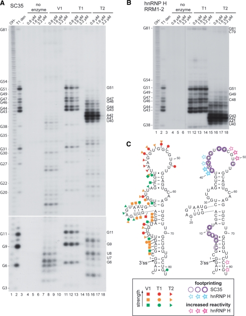
Alternative splicing is regulated in part by variations in the relative concentrations of a variety of factors, including serine/arginine-rich (SR) proteins. The SR protein SC35 self-regulates its expression by stimulating unproductive splicing events in the 3' untranslated region of its own pre-mRNA. Using various minigene constructs containing the terminal retained intron and flanking exons, we identified in the highly conserved last exon a number of exonic splicing enhancer elements responding specifically to SC35, and showed an inverse correlation between affinity of SC35 and enhancer strength. The enhancer region, which is included in a long stem loop, also contains repressor elements, and is recognized by other RNA-binding proteins, notably hnRNP H protein and TAR DNA binding protein (TDP-43). Finally, in vitro and in cellulo experiments indicated that hnRNP H and TDP-43 antagonize the binding of SC35 to the terminal exon and specifically repress the use of SC35 terminal 3' splice site. Our study provides new information about the molecular mechanisms of SC35-mediated splicing activation. It also highlights the existence of a complex network of self- and cross-regulatory mechanisms between splicing regulators, which controls their homeostasis and offers many ways of modulating their concentration in response to the cellular environment.


Figures

Figure 1.
Figure 1.
(A) Schematic organization of the SC35 gene (SFRS2) and of the SC35-RI minigene. The open-reading frame lies within two constitutive exons, whereas the 3′ UTR is alternatively spliced and polyadenylated. The alternative cassette exon and retained intron are represented by black boxes. (B) Sequence of the region encompassing the 3′ splice site of terminal intron and the 5′ part of terminal exon. Intronic and exonic nucleotides are in small and capital letters, respectively. Numbers along the exonic sequence indicate the position relative to the 3′ splice site in the wild-type construct. In the wt* construct, the three newly created restriction sites are underlined. The different mutated fragments are in bold. Note that they do not systematically have the same length as the corresponding wild-type fragment. (C) In vitro splicing of wild-type and mutant SC35-RI transcripts. Standard splicing assays were carried out in HeLa nuclear extract, supplemented or not by 600–800 ng of purified full-length SC35, ASF/SF2 or the same proteins lacking the RS domain (ΔRS). The different pre-mRNAs are indicated above each panel. The pre-mRNA and splicing products are symbolized on the side of the gel. Products resulting from the use of an intronic cryptic 5′ splice site (intronic position +157) or from the use of an exonic cryptic 3′ splice site are indicated by the mention ‘cryptic’ and by an asterisk, respectively. The activity of SC35 and ASF/SF2 proteins was normalized using a control fushi-tarazu transcript (Supplementary Figure S1A).
Figure 2.
Figure 2.
(A) Schematic view of the heterologous Sp1 ‘inverted exon 2’ splicing substrate. Various sequences, as listed on the right side of the figure, were inserted in the middle of the inverted exon 2. Like for sequences from the terminal SC35 exon, the fragments 1, 2 and 3 from the cassette exon partially overlap one to another (underlined nts). (B) Splicing assays in nuclear extract. Each transcript, harbouring one specific sequence in exon 2 as indicated, was incubated and analysed in standard conditions. (C) Splicing assays in cytoplasmic S100 extract. Transcripts, containing either the S-94 or the IIB sequence, were incubated in 8 µl S100 supplemented with 3 µl of a 20–40% ammonium sulphate-precipitated fraction from HeLa nuclear extract and with 400 ng of recombinant SC35 or ASF/SF2 protein. As previously shown (25,30), efficient and specific splicing activation in the S100 fraction could only be observed in the presence of this 20–40% fraction, which does not contain SR proteins.
Figure 3.
Figure 3.
(A) Electrophoretic mobility shift assay. The complexes formed between the RNA probes (10 fmol) and GST-SC35ΔRS (10, 20 and 30 ng) or GST-9G8ΔRS (30 ng) were resolved by native gel electrophoresis to separate the protein-RNA complexes (bound) from the free RNA (free). (B) Sequence homology between the sequence IIB from the SC35 terminal exon (nts 46–65) and SC35 high affinity sequences S-7 and S-94 sequences. (C) Western-blot of an RNA affinity experiment. Equivalent amounts of proteins retained on the different immobilized RNA probes (lanes 1 to 4), or on naked beads (lane 5) were analysed using various antibodies. The input corresponded to 1.5 µl of HeLa nuclear extract. nd: not determined.
Figure 4.
Figure 4.
(A) The RNA was incubated in the absence (−) or in the presence of different concentrations of SC35 recombinant protein, as indicated above each lane. Digestions with RNases V1 (lanes 7–10), T1 (lanes 11–14) or T2 (lanes 15–18) were carried out as described in the ‘Materials and methods’ section. As a control, undigested RNA was fractionated in parallel (lanes 3–6). Lanes OH− and T1 den, corresponding respectively to alkaline hydrolysis and RNase T1 digestion in denaturing conditions, were used for localization of the cleavage sites. Nucleotides with decreased sensitivity to RNases in the presence of SC35 protein are indicated on the right. (B) The same experiment as in A was done using the hnRNP H RRM1-2 recombinant protein. (C) Secondary structure model proposed for SC35 terminal exon. The model was proposed based on thermodynamic considerations and on the results of enzymatic digestions shown in A and B. V1, T1 and T2 RNase cleavages are represented by arrows surmounted by squares, dots and triangles, respectively. Red, orange and green symbols indicate a strong, medium or low cleavage, respectively. The residues protected by SC35 (1.6 µM) or hnRNP H RRM1-2 (3.2 µM), or having a modified sensitivity in the presence of hnRNP H RRM1-2 (3.2 µM) are circled in purple or indicated by a blue or a pink star, respectively (the line thickness is proportional to the protection strength).
Figure 5.
Figure 5.
UV cross-linking assays. RNA probes I (lanes 1–8), IIB (lanes 9–16), S-7 (lanes 17–20) or control C (lanes 21–24) (see sequences in Figure 2A) were incubated with 2 µl of cytoplasmic extract S100 alone (lanes 1, 9, 17 and 21) or complemented with the following concentrations of purified recombinant proteins, as indicated above the figure: 400 nM FLAG-SC35, 250 and 500 nM hnRNP H, 175 and 350 nM TDP-43 (or the maximal amount of either protein added in lanes 7–8 and 15–16). Both recombinant hnRNP H and TDP-43 proteins migrated as double bands, which resulted either from premature termination of translation or from partial proteolytic digestion during the purification procedure (data not shown). The protein marked by an asterisk, which cross-linked only to the S-7 RNA in the S100 extract (lane 17), might correspond to residual amount of endogenous SC35 that escaped the nuclei during the preparation of the extract.
Figure 6.
Figure 6.
(A) Repression of SC35-RI pre-mRNA splicing by hnRNP proteins. The transcript is schematized on top of the figure. Splicing was carried out in standard conditions in the absence or presence of 300, 320 or 480 ng of purified recombinant GST-hnRNP H, hnRNP A1 or GST-TDP-43, respectively, or half of those amounts when two proteins were added together. Splicing efficiencies were calculated as in Supplementary Figure S5 and are indicated under each lane. (B) Splicing of Sp1-derived pre-mRNA in the presence of recombinant hnRNP proteins. The different transcripts are schematized on top of each panel. The common backbone derived from the wild-type Sp1 pre-mRNA (top right panel) is represented by grey boxes. The white box in the chimeric Sp1-ter-ex transcript symbolizes the terminal exon from the SC35-RI transcript, while the minimal SC35 IIB sequence and the control ASF/SF2-specific sequence are represented by a small white (Sp1-IIB) or black (Sp1-ASF) box, respectively. The experiment was carried out as in A.
Figure 7.
Figure 7.
(A) Schematic structure of the pSC35-βGlo minigene. A fragment of the SC35 gene including the 3′ part of the retained terminal intron (thick black line) and the 5′ part of the terminal exon (white box) was inserted in the rabbit β-globin intron 2, creating a reporter with alternative 3′ splice sites (splicing is represented by broken filled lines). When the SC35 3′ splice site was used, a cryptic 5′ splice site located downstream from the insert, within the 3′ part of the β-globin intron, was systematically activated (broken dotted line), creating an ‘SC35’ alternative exon. (B) and (C) RNA and protein analysis after transfection of HeLa cells. Cells were cotransfected with the pSC35-βGlo minigene and plasmids carrying various cDNAs as indicated above the gels. RNA and protein samples were extracted and analysed respectively by RT-PCR (B) or western blotting (C) with specific antibodies, as indicated. Anti-GFP was used to monitor the amount of GFP-SC35 protein, which could not be detected using anti-SC35 antibodies due to its low level of expression. AS gave rise to two major mRNA products depending on the use of the β-globin (lower band) or SC35 (upper band) 3′ splice site. (D) and (E) RNA and protein analysis after siRNA-mediated knockdown of hnRNP H/F or TDP-43. RT-PCR analysis and quantification of mRNA products (D) was as in panel B. Efficiency of silencing and endogenous level of SC35 protein were monitored by western-blotting (E) as in panel C.

Similar articles

Cited by

References

    1. Wang Z, Burge CB. Splicing regulation: from a parts list of regulatory elements to an integrated splicing code. RNA. 2008;14:802–813. - PMC - PubMed
    1. Bourgeois CF, Lejeune F, Stevenin J. Broad specificity of SR (serine/arginine) proteins in the regulation of alternative splicing of pre-messenger RNA. Prog. Nucleic Acid Res. Mol. Biol. 2004;78:37–88. - PubMed
    1. Long JC, Caceres JF. The SR protein family of splicing factors: master regulators of gene expression. Biochem. J. 2009;417:15–27. - PubMed
    1. Martinez-Contreras R, Cloutier P, Shkreta L, Fisette JF, Revil T, Chabot B. hnRNP proteins and splicing control. Adv. Exp. Med. Biol. 2007;623:123–147. - PubMed
    1. Pandit S, Wang D, Fu XD. Functional integration of transcriptional and RNA processing machineries. Curr. Opin. Cell Biol. 2008;20:260–265. - PMC - PubMed

Publication types

MeSH terms

Substances