. 2010 Feb 15;181(4):344-52.
doi: 10.1164/rccm.200906-0826OC. Epub 2009 Dec 3.

Stimulation of sphingosine 1-phosphate signaling as an alveolar cell survival strategy in emphysema

Affiliations

Stimulation of sphingosine 1-phosphate signaling as an alveolar cell survival strategy in emphysema

Khalil J Diab et al. Am J Respir Crit Care Med. .

Abstract

Rationale: Vascular endothelial growth factor receptor (VEGFR) inhibition increases ceramides in lung structural cells of the alveolus, initiating apoptosis and alveolar destruction morphologically resembling emphysema. The effects of increased endogenous ceramides could be offset by sphingosine 1-phosphate (S1P), a prosurvival by-product of ceramide metabolism.

Objectives: The aims of our work were to investigate the sphingosine-S1P-S1P receptor axis in the VEGFR inhibition model of emphysema and to determine whether stimulation of S1P signaling is sufficient to functionally antagonize alveolar space enlargement.

Methods: Concurrent to VEGFR blockade in mice, S1P signaling augmentation was achieved via treatment with the S1P precursor sphingosine, S1P agonist FTY720, or S1P receptor-1 (S1PR1) agonist SEW2871. Outcomes included sphingosine kinase-1 RNA expression and activity, sphingolipid measurements by combined liquid chromatography-tandem mass spectrometry, immunoblotting for prosurvival signaling pathways, caspase-3 activity and terminal deoxynucleotidyltransferase-mediated dUTP nick end labeling assays, and airspace morphometry.

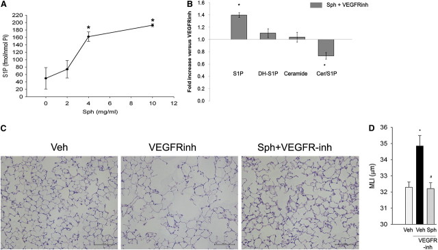
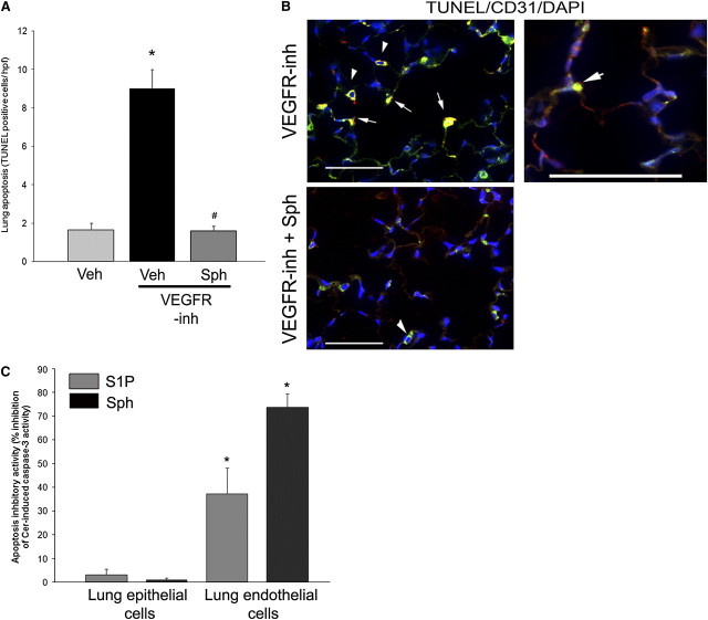
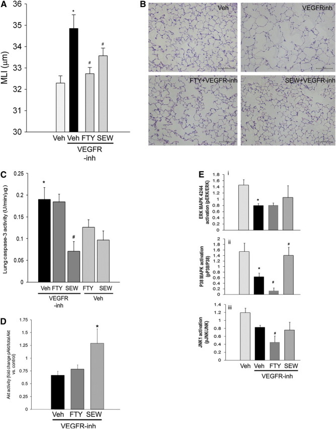
Measurements and main results: Consistent with previously reported de novo activation of ceramide synthesis, VEGFR inhibition triggered increases in lung ceramides, dihydroceramides, and dihydrosphingosine, but did not alter sphingosine kinase activity or S1P levels. Administration of sphingosine decreased the ceramide-to-S1P ratio in the lung and inhibited alveolar space enlargement, along with activation of prosurvival signaling pathways and decreased lung parenchyma cell apoptosis. Sphingosine significantly opposed ceramide-induced apoptosis in cultured lung endothelial cells, but not epithelial cells. FTY720 or SEW2871 recapitulated the protective effects of sphingosine on airspace enlargement concomitant with attenuation of VEGFR inhibitor-induced lung apoptosis.

Conclusions: Strategies aimed at augmenting the S1P-S1PR1 signaling may be effective in ameliorating the apoptotic mechanisms of emphysema development.


Figures

Figure 1.
Figure 1.
(AF) Ceramide and sphingosine 1-phosphate (S1P) expression in response to vascular endothelial growth factor receptor (VEGFR) inhibition. Sphingolipid expression (mean + SEM; n = 3–6) in the lung after administration of vehicle (Veh; carboxymethylcellulose, 0.5%) or the VEGFR inhibitor SU5416 (VEGFR-inh, 20 mg/kg; Day 7): (A) S1P levels, normalized by lipid inorganic phosphorus (Pi); (B) dihydro-S1P levels, P = 0.03; (C) ceramide levels, P = 0.001; (D) dihydro-ceramide lung levels, P = 0.001; (E) [33P]SPHK-1 activity in the lung; (F) ratio of ceramide to S1P in the lung, P = 0.02.
Figure 2.
Figure 2.
Effect of d-sphingosine supplementation on vascular endothelial growth factor receptor (VEGFR) inhibitor–induced airspace enlargement. (A) Sphingosine 1-phosphate (S1P) levels in the lung on Day 5 after administration of sphingosine (Sph) at the indicated concentrations (mean ± SEM; *P < 0.05; n = 2 or 3 per time point). (B) Changes in sphingolipids S1P, dihydro-S1P (DH-S1P), ceramide, and the ratio of ceramide to S1P in the lungs of mice receiving sphingosine supplementation after VEGFR inhibitor (VEGFR-inh) administration, compared with those animals that were treated with VEGFR inhibitor with vehicle (mean ± SEM; *P < 0.05; n = 5). (C) Representative micrographs of hematoxylin and eosin–stained sections of inflated and fixed alveolar lung tissue from mice treated with vehicle (Veh; 0.5% carboxymethylcellulose), VEGFR-inh (SU5416, 20 mg/kg, subcutaneous; Day 28), or d-sphingosine (Sph; 20 μg, intraperitoneal, daily on Days 1–5 after SU5416 administration). Scale bars, 100 μm. Note the airspace enlargement in the VEGFR inhibitor–treated lungs, which is reduced by treatment with Sph. (D) Morphometric measurements of the mean linear intercept (MLI), indicating airspace size in mice that were treated with Veh, VEGFR-inh, or Sph and VEGFR-inh (mean + SEM; *P < 0.05 vs. Veh; #P < 0.05 vs. VEGFR-inh; n = 5 per group; analysis of variance).
Figure 3.
Figure 3.
Effect of vascular endothelial growth factor receptor (VEGFR) inhibition and d-sphingosine supplementation on Akt and mitogen-activated protein kinase (MAPK) signaling pathways in the lung. Protein homogenates were isolated 28 days after administration of the VEGFR inhibitor SU5416 (VEGFR-inh; 20 mg/kg, once), with concomitant d-sphingosine (Sph; 20 μg/injection for 5 d) or its vehicle (Veh) and protein was used for immunoblotting (30 μg) with the respective antibodies, followed by densitometry. Each lane represents an individual animal. (A) Akt activation was measured as the ratio of phosphorylated Akt to total Akt, as detected by immunoblotting (right) (mean + SEM; *P < 0.05 vs. Veh; n = 4 or 5). (B) Extracellular signal–regulated kinase (ERK) (MAPK 42/44) activation was measured as the ratio of phosphorylated ERK to total ERK, as detected by immunoblotting (right) (mean + SEM; *P < 0.05 vs. Veh; #P < 0.05 vs. VEGFR-inh; analysis of variance [ANOVA]; n = 4–8). (C) p38 MAPK activation was measured as the ratio of phosphorylated p38 MAPK to total p38 MAPK, as detected by immunoblotting (right) (mean + SEM; *P < 0.05 vs. Veh; #P < 0.05 vs. VEGFR-inh; ANOVA; n = 4–8). (D) c-Jun N-terminal kinase-1 (JNK1) activation was measured as the ratio of phosphorylated JNK1 to total JNK1 (mean + SEM; *P < 0.05 vs. Veh; ANOVA; n = 3–8).
Figure 4.
Figure 4.
Antiapoptotic effects of d-sphingosine. (A) Lung apoptosis (number of terminal deoxynucleotidyltransferase–mediated dUTP nick end labeling [TUNEL]-positive cells per high-power lung field [hpf] measured on coded slides) was increased in response to vascular endothelial growth factor receptor inhibitor (VEGFR-inh), but not in mice supplemented with d-sphingosine (Sph) (mean + SEM; *P < 0.05 vs. vehicle [Veh]; #P < 0.05 vs. VEGFR-inh; Student's t test; n = 3–6). (B) Coimmunolocalization of TUNEL-positive apoptotic cells (green) with endothelial cell marker (CD31, red) in lungs treated with VEGFR-inh with or without Sph supplementation. Note TUNEL-positive nuclear staining of CD-31 decorated cells (arrows). Alveolar macrophages exhibited typical green autofluorescence (arrowheads). Scale bars, 50 μm. (C) Inhibitory activity of sphingosine 1-phosphate (S1P, 1 μM) or Sph (1 μM) cotreatments on ceramide-induced caspase-3 activity measured by kinetic fluorimetry (U/min) normalized by protein concentration in lysates of primary rat lung epithelial and endothelial cells. The inhibitory activity was calculated relative to the apoptotic activity induced by ceramide 16:0 (Cer, 10 μM, 24 h) treatment in the absence of S1P or Sph (mean + SEM; *P < 0.05 vs. inhibitory activity of the respective S1P agonist on epithelial cells; n = 3). DAPI = 4′,6-diamidino-2-phenylindole.
Figure 5.
Figure 5.
Effects of sphingosine 1-phosphate (S1P) receptor agonists on vascular endothelial growth factor receptor (VEGFR) blockade–induced airspace enlargement, apoptosis, and signaling. After administration of VEGFR inhibitor (VEGFR-inh) SU5416, with or without concomitant S1P receptor agonist FTY720 (0.1 mg/kg/day, daily, intraperitoneal, administered on Days 1–5 after SU5416) or SEW2871 (20 mg/kg, daily by gavage, administered on Days 1–5 after SU5416), airspace enlargement was quantified by mean linear intercept (MLI; Day 28 [A]) (mean + SEM, *P < 0.05 vs. control, #P < 0.05 vs. VEGFR-inh; n = 5). (B) Representative panels of hematoxylin and eosin–stained lung parenchyma. Scale bars, 100 μm. (C) Lung caspase-3 activity measured by kinetic fluorimetry in total lung lysates 28 days after VEGFR inhibitor administration. Note that SEW2871 significantly decreased VEGFR inhibitor–induced lung caspase-3 activity (mean + SEM; *P < 0.05 vs. control, #P < 0.05 vs. VEGFR-inh; n = 3–5). (D) Changes in lung Akt phosphorylation compared with controls measured by densitometry 28 days after VEGFR inhibitor administration (mean fold change vs. carboxymethylcellulose [CMC] control + SEM; #P < 0.05 vs. VEGFR-inh). (E) Mitogen-activated protein kinase (MAPK) activity in tissue protein (30 μg) immunoblotted with the respective antibodies and measured by densitometry of phospho- vs. total extracellular signal–regulated kinase (ERK; panel i), p38 MAPK (panel ii), or c-Jun N-terminal kinase-1 (JNK1) (panel iii).

Similar articles

Cited by

References

    1. National Heart, Lung, and Blood Institute. The definition of emphysema: report of a National Heart, Lung, and Blood Institute, Division of Lung Diseases Workshop. Am Rev Respir Dis 1985;132:182–185. - PubMed
    1. Tuder RM, Petrache I, Elias JA, Voelkel NF, Henson PM. Apoptosis and emphysema: the missing link. Am J Respir Cell Mol Biol 2003;28:551–554. - PubMed
    1. Giordano RJ, Lahdenranta J, Zhen L, Chukwueke U, Petrache I, Langley RR, Fidler IJ, Pasqualini R, Tuder RM, Arap W. Targeted induction of lung endothelial cell apoptosis causes emphysema-like changes in the mouse. J Biol Chem 2008;283:29447–29460. - PMC - PubMed
    1. Aoshiba K, Yokohori N, Nagai A. Alveolar wall apoptosis causes lung destruction and emphysematous changes. Am J Respir Cell Mol Biol 2003;28:555–562. - PubMed
    1. Kasahara Y, Tuder RM, Taraseviciene-Stewart L, Le Cras TD, Abman S, Hirth PK, Waltenberger J, Voelkel NF. Inhibition of VEGF receptors causes lung cell apoptosis and emphysema. J Clin Invest 2000;106:1311–1319. - PMC - PubMed

MeSH terms