

Extracellular production of an RNA aptamer by ribonuclease-free marine bacteria harboring engineered plasmids: a proposal for industrial RNA drug production
- PMID: 19966026
- PMCID: PMC2813014
- DOI: 10.1128/AEM.01971-09
Extracellular production of an RNA aptamer by ribonuclease-free marine bacteria harboring engineered plasmids: a proposal for industrial RNA drug production
Abstract
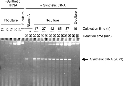
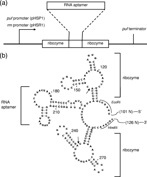
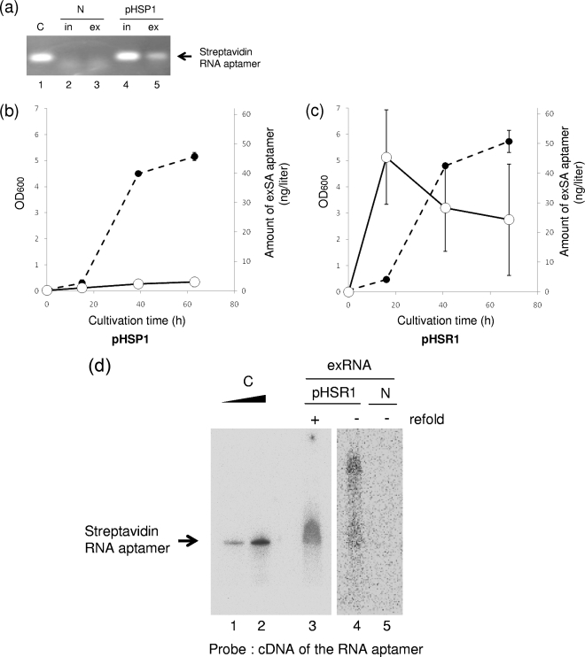
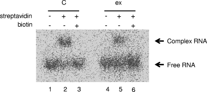
Natural noncoding small RNAs have been shown to be involved in a number of cellular processes as regulators. Using the mechanisms thus elucidated, artificial small interfering RNAs (siRNAs), ribozymes, and RNA aptamers are also expected to be potential candidates for RNA therapeutic agents. However, current techniques are too costly for industrial production of these RNAs for use as drugs. Here, we propose a new method for in vivo production of artificial RNAs using the marine phototrophic bacterium Rhodovulum sulfidophilum. Using engineered plasmids and this bacterium, which produces extracellular nucleic acids in nature, we developed a method for extracellular production of a streptavidin RNA aptamer. As the bacterium does not produce any RNases in the culture medium, at least within the cultivation period tested, the designed RNA itself is produced and retained in the culture medium of the bacterium without any specific mechanism for protection against degradation by nucleases. Here, we report that the streptavidin RNA aptamer is produced in the culture medium and retains its specific function. This is the first demonstration of extracellular production of a functional artificial RNA in vivo, which will pave the way for inexpensive production of RNA drugs.
Figures
Similar articles
-
Artificial RNA aptamer production by the marine bacterium Rhodovulum sulfidophilum: improvement of the aptamer yield using a mutated transcriptional promoter.J Biosci Bioeng. 2011 Nov;112(5):458-61. doi: 10.1016/j.jbiosc.2011.07.025. Epub 2011 Sep 8. J Biosci Bioeng. 2011. PMID: 21903467
-
Extracellular nucleic acids of the marine bacterium Rhodovulum sulfidophilum and recombinant RNA production technology using bacteria.FEMS Microbiol Lett. 2018 Feb 1;365(3). doi: 10.1093/femsle/fnx268. FEMS Microbiol Lett. 2018. PMID: 29228187 Review.
-
Short hairpin RNAs of designed sequences can be extracellularly produced by the marine bacterium Rhodovulum sulfidophilum.J Gen Appl Microbiol. 2014;60(6):222-6. doi: 10.2323/jgam.60.222. J Gen Appl Microbiol. 2014. PMID: 25742972
-
Characterization of extracellular DNA production and flocculation of the marine photosynthetic bacterium Rhodovulum sulfidophilum.Appl Microbiol Biotechnol. 2009 Aug;84(2):349-56. doi: 10.1007/s00253-009-2031-7. Epub 2009 May 19. Appl Microbiol Biotechnol. 2009. PMID: 19452150
-
RNases in ColE1 DNA metabolism.Mol Biol Rep. 1995-1996;22(2-3):195-200. doi: 10.1007/BF00988728. Mol Biol Rep. 1995. PMID: 8901510 Review.
Cited by
-
Engineering nucleic acid structures for programmable molecular circuitry and intracellular biocomputation.Nat Chem. 2017 Nov;9(11):1056-1067. doi: 10.1038/nchem.2852. Epub 2017 Sep 25. Nat Chem. 2017. PMID: 29064489 Free PMC article. Review.
-
A guide to large-scale RNA sample preparation.Anal Bioanal Chem. 2018 May;410(14):3239-3252. doi: 10.1007/s00216-018-0943-8. Epub 2018 Mar 15. Anal Bioanal Chem. 2018. PMID: 29546546 Free PMC article. Review.
-
Novel approaches for efficient in vivo fermentation production of noncoding RNAs.Appl Microbiol Biotechnol. 2020 Mar;104(5):1927-1937. doi: 10.1007/s00253-020-10350-3. Epub 2020 Jan 17. Appl Microbiol Biotechnol. 2020. PMID: 31953559 Free PMC article. Review.
-
RNAi-Based Therapeutics and Novel RNA Bioengineering Technologies.J Pharmacol Exp Ther. 2023 Jan;384(1):133-154. doi: 10.1124/jpet.122.001234. Epub 2022 Jun 9. J Pharmacol Exp Ther. 2023. PMID: 35680378 Free PMC article. Review.
-
Marine Purple Photosynthetic Bacteria as Sustainable Microbial Production Hosts.Front Bioeng Biotechnol. 2019 Oct 11;7:258. doi: 10.3389/fbioe.2019.00258. eCollection 2019. Front Bioeng Biotechnol. 2019. PMID: 31681740 Free PMC article. Review.
References
-
- Ando, T., H. Suzuki, S. Nishimura, T. Tanaka, A. Hiraishi, and Y. Kikuchi. 2006. Characterization of extracellular RNAs produced by the marine photosynthetic bacterium Rhodovulum sulfidophilum. J. Biochem. 139:805-811. - PubMed
-
- Breaker, R. R. 2004. Natural and engineered nucleic acids as tools to explore biology. Nature 432:838-845. - PubMed
-
- Chu, C. Y., and T. M. Rana. 2007. Small RNAs: regulators and guardians of the genome. J. Cell Physiol. 213:412-419. - PubMed
Publication types
MeSH terms
Substances
LinkOut - more resources
Full Text Sources
Other Literature Sources
Molecular Biology Databases
Research Materials
