Comparative Study
. 2010 Jan;89(1):77-81.
doi: 10.1177/0022034509352151.

Bone regeneration in defects compromised by radiotherapy

Affiliations
Comparative Study

Bone regeneration in defects compromised by radiotherapy

W-W Hu et al. J Dent Res. 2010 Jan.

Abstract

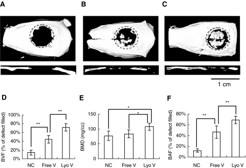
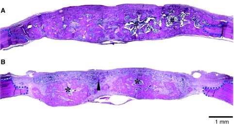
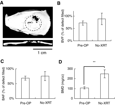
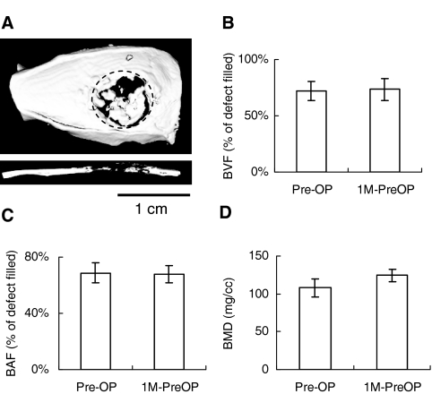
Because bone reconstruction in irradiated sites is less than ideal, we applied a regenerative gene therapy method in which a cell-signaling virus was localized to biomaterial scaffolds to regenerate wounds compromised by radiation therapy. Critical-sized defects were created in rat calvariae previously treated with radiation. Gelatin scaffolds containing lyophilized adenovirus encoding BMP-2 (AdBMP-2) or freely suspended AdBMP-2 were transplanted. Lyophilized AdBMP-2 significantly improved bone quality and quantity over free AdBMP-2. Bone mineral density was reduced after radiotherapy. Histological analyses demonstrated that radiation damage led to less bone regeneration. The woven bone and immature marrow formed in the radiated defects indicated that irradiation retarded normal bone development. Finally, we stored the scaffolds with lyophilized AdBMP-2 at -80 degrees C to determine adenovirus stability. Micro-CT quantification demonstrated no significant differences between bone regeneration treated with lyophilized AdBMP-2 before and after storage, suggesting that virus-loaded scaffolds may be convenient for application as pre-made constructs.


Figures

Figure 1.
Figure 1.
Bone formation in critical-sized calvarial defects compromised by radiation therapy. We performed µ-CT analysis to visualize and quantify bone regeneration. The 3-D images were reconstructed to illustrate the top and sagittal section views of (A) AdLacZ lyophilized in gelatin scaffolds, (B) AdBMP-2 freely suspended in gelatin scaffolds, and (C) AdBMP-2 lyophilized in gelatin scaffolds. The newly formed bone was evaluated by (D) bone volume fraction (BVF) in defects, by (E) bone mineral density (BMD) of newly formed bone, and by (F) bone area fraction (BAF) in defects assessed from the projected area ratio of the µ-CT image. The data were expressed as mean ± standard deviation, and the statistical differences between groups were analyzed by Student's t test (*p < 0.05; **p < 0.01). The dotted line represents the surgical margin of the cranial defect. N = 5.
Figure 2.
Figure 2.
Histological analysis of critical-sized calvarial defects with or without pre-operative radiation therapy. Sections were prepared from the midline of defects. The defects without (A) and with (B) radiation treatment were examined. The defect margins are depicted by blue dashed lines. Arrowhead, non-degraded gelatin sponges; #mature bone marrow with adipose tissues; *immature bone marrow.
Figure 3.
Figure 3.
Bone formation in critical-sized calvarial defects without radiation therapy (No-XRT). (A) The 3-D images were reconstructed to illustrate the top and sagittal section views. Bone regeneration was compared with that in the irradiated group (Pre-OP) by µ-CT analyses: (B) BVF, (C) BAF, and (D) BMD. The data were expressed as mean ± standard deviation, and the statistical differences between groups were analyzed by Student's t test (*p < 0.05; **p < 0.01). The dotted line represents the surgical margin of the cranial defect. N = 5.
Figure 4.
Figure 4.
Irradiated defects regenerated with AdBMP-2 lyophilized in gelatin scaffolds. To test the stability of viruses in the gelatin scaffolds, we stored pre-made AdBMP-2-lyophilized scaffolds (1M-PreOP) at -80°C for 1 mo prior to surgery. (A) The 3-D images were reconstructed to illustrate the top and sagittal section views. The bone regeneration was compared with that in the radiated group before storage (Pre-OP) by µ-CT analyses: (B) BVF, (C) BAF, and (D) BMD. The data were expressed as mean ± standard deviation, and the statistical differences between groups were analyzed by Student's t test (*p < 0.05; **p < 0.01). The dotted line represents the surgical margin of the cranial defect. N = 5.

References

    1. Chang SC, Wei FC, Chuang HL, Chen YR, Chen JK, Lee KC, et al. (2003). Ex vivo gene therapy in autologous critical-size craniofacial bone regeneration. Plast Reconstr Surg 112:1841-1850 - PubMed
    1. Deutsch M, Kroll SS, Ainsle N, Wang B. (1999). Influence of radiation on late complications in patients with free fibular flaps for mandibular reconstruction. Ann Plast Surg 42:662-664 - PubMed
    1. Disa JJ, Cordeiro PG. (2000). Mandible reconstruction with microvascular surgery. Semin Surg Oncol 19:226-234 - PubMed
    1. Ehrhart NP, Hong L, Morgan AL, Eurell JA, Jamison RD. (2005). Effect of transforming growth factor-beta1 on bone regeneration in critical-sized bone defects after irradiation of host tissues. Am J Vet Res 66:1039-1045 - PubMed
    1. Emerick KS, Teknos TN. (2007). State-of-the-art mandible reconstruction using revascularized free-tissue transfer. Expert Rev Anticancer Ther 7:1781-1788 - PubMed

Publication types

MeSH terms