Reduced expression of Cx43 attenuates ventricular remodeling after myocardial infarction via impaired TGF-beta signaling
- PMID: 19966054
- PMCID: PMC2822575
- DOI: 10.1152/ajpheart.00806.2009
Reduced expression of Cx43 attenuates ventricular remodeling after myocardial infarction via impaired TGF-beta signaling
Abstract
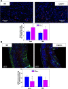
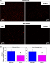
In addition to mediating cell-to-cell electrical coupling, gap junctions are important in tissue repair, wound healing, and scar formation. The expression and distribution of connexin43 (Cx43), the major gap junction protein expressed in the heart, are altered substantially after myocardial infarction (MI); however, the effects of Cx43 remodeling on wound healing and the attendant ventricular dysfunction are incompletely understood. Cx43-deficient and wild-type mice were subjected to proximal ligation of the anterior descending coronary artery and followed for 6 days or 4 wk to test the hypothesis that reduced expression of Cx43 influences wound healing, fibrosis, and ventricular remodeling after MI. We quantified the progression of infarct healing by measuring neutrophil expression, collagen content, and myofibroblast expression. We found significantly reduced transformation of fibroblasts to myofibroblasts at 6 days and significantly reduced collagen deposition both in the infarct at 6 days and at 4 wk in the noninfarcted region of Cx43-deficient mice. As expected, transforming growth factor (TGF)-beta, a profibrotic cytokine, was dramatically upregulated in MI hearts, but its phosphorylated comediator (pSmad) was significantly downregulated in the nuclei of Cx43-deficient hearts post-MI, suggesting that downstream signaling of TGF-beta is diminished substantially in Cx43-deficient hearts. This diminution in profibrotic TGF-beta signaling resulted in the attenuation of adverse structural remodeling as assessed by echocardiography. These findings suggest that efforts to enhance the expression of Cx43 to maintain intercellular coupling or reduce susceptibility to arrhythmias should be met with caution until the role of Cx43 in infarct healing is fully understood.
Figures


Similar articles
-
Remodeling of cardiac fibroblasts following myocardial infarction results in increased gap junction intercellular communication.Cardiovasc Pathol. 2010 Nov-Dec;19(6):e233-40. doi: 10.1016/j.carpath.2009.12.002. Epub 2010 Jan 25. Cardiovasc Pathol. 2010. PMID: 20093048 Free PMC article.
-
Connexin43 as a determinant of myocardial infarct size following coronary occlusion in mice.J Am Coll Cardiol. 2003 Feb 19;41(4):681-6. doi: 10.1016/s0735-1097(02)02893-0. J Am Coll Cardiol. 2003. PMID: 12598083
-
MK5 haplodeficiency decreases collagen deposition and scar size during post-myocardial infarction wound repair.Am J Physiol Heart Circ Physiol. 2019 Jun 1;316(6):H1281-H1296. doi: 10.1152/ajpheart.00532.2017. Epub 2019 Mar 22. Am J Physiol Heart Circ Physiol. 2019. PMID: 30901279 Free PMC article.
-
The role of TGF-beta signaling in myocardial infarction and cardiac remodeling.Cardiovasc Res. 2007 May 1;74(2):184-95. doi: 10.1016/j.cardiores.2006.10.002. Epub 2006 Oct 7. Cardiovasc Res. 2007. PMID: 17109837 Free PMC article. Review.
-
Post translational modifications of connexin 43 in ventricular arrhythmias after myocardial infarction.Mol Biol Rep. 2024 Feb 23;51(1):329. doi: 10.1007/s11033-024-09290-2. Mol Biol Rep. 2024. PMID: 38393658 Review.
Cited by
-
Canagliflozin Improves Myocardial Perfusion, Fibrosis, and Function in a Swine Model of Chronic Myocardial Ischemia.J Am Heart Assoc. 2023 Jan 3;12(1):e028623. doi: 10.1161/JAHA.122.028623. Epub 2022 Dec 30. J Am Heart Assoc. 2023. PMID: 36583437 Free PMC article.
-
Role of Non-Myocyte Gap Junctions and Connexin Hemichannels in Cardiovascular Health and Disease: Novel Therapeutic Targets?Int J Mol Sci. 2018 Mar 15;19(3):866. doi: 10.3390/ijms19030866. Int J Mol Sci. 2018. PMID: 29543751 Free PMC article. Review.
-
Myofibroblasts impair myocardial impulse propagation by heterocellular connexin43 gap-junctional coupling through micropores.Front Physiol. 2024 Feb 23;15:1352911. doi: 10.3389/fphys.2024.1352911. eCollection 2024. Front Physiol. 2024. PMID: 38465264 Free PMC article.
-
Sympatho-adrenergic mechanisms in heart failure: new insights into pathophysiology.Med Rev (2021). 2021 Oct 21;1(1):47-77. doi: 10.1515/mr-2021-0007. eCollection 2021 Oct. Med Rev (2021). 2021. PMID: 37724075 Free PMC article.
-
Reduced connexin-43 expression, slow conduction and repolarisation dispersion in a model of hypertrophic cardiomyopathy.Dis Model Mech. 2024 Aug 1;17(8):dmm050407. doi: 10.1242/dmm.050407. Epub 2024 Aug 27. Dis Model Mech. 2024. PMID: 39189070 Free PMC article.
References
-
- Asazuma-Nakamura Y, Dai P, Harada Y, Jiang Y, Hamaoka K, Takamatsu T. Cx43 contributes to TGF-β signaling to regulate differentiation of cardiac fibroblasts into myofibroblasts. Exp Cell Res 315: 1190–1199, 2009. - PubMed
-
- Bajpai S, Shukla VK, Tripathi K, Srikrishna S, Singh RK. Targeting connexin 43 in diabetic wound healing: future perspectives. J Postgrad Med 55: 143–149, 2009. - PubMed
-
- Banerjee I, Yekkala K, Borg TK, Baudino TA. Dynamic interactions between myocytes, fibroblasts, and extracellular matrix. Ann NY Acad Sci 1080: 76–84, 2006. - PubMed
-
- Betsuyaku T, Kovacs A, Saffitz JE, Yamada KA. Cardiac structure and function in young and senescent mice heterozygous for a connexin43 null mutation. J Mol Cell Cardiol 34: 175–184, 2002. - PubMed
Publication types
MeSH terms
Substances
Grants and funding
LinkOut - more resources
Full Text Sources
Medical
Molecular Biology Databases
Research Materials
Miscellaneous

