. 2010 Feb;151(2):731-40.
doi: 10.1210/en.2009-0955. Epub 2009 Dec 4.

Epigenetic regulation of estrogen receptor alpha gene expression in the mouse cortex during early postnatal development

Affiliations

Epigenetic regulation of estrogen receptor alpha gene expression in the mouse cortex during early postnatal development

Jenne M Westberry et al. Endocrinology. 2010 Feb.

Abstract

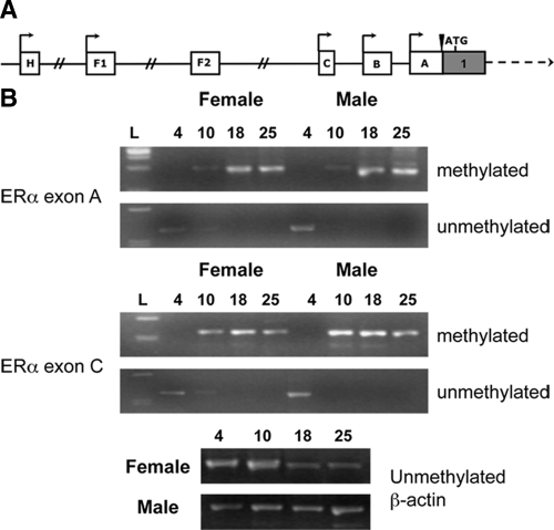
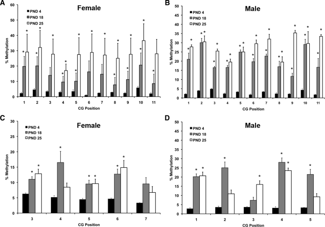
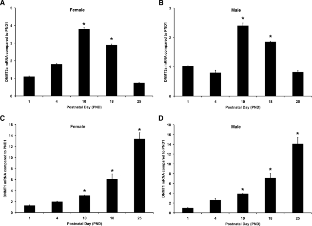
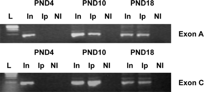
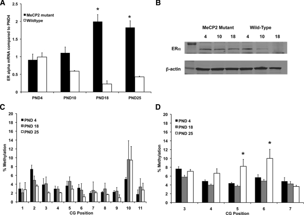
Estrogens play a critical role in brain development by acting on areas that express estrogen receptors. In the rodent cortex, estrogen receptor alpha (ER alpha) mRNA expression is high early in postnatal development but declines starting at postnatal day (PND) 10 and is virtually absent in the adult cortex. The mechanisms controlling this regulation are largely unknown. Methylation is important for gene silencing during development in many tissues, including the brain. In the present study, we examined the methylation status of ER alpha 5' untranslated exons during early postnatal development in male and female mice using methylation-specific PCR and pyrosequencing. Several regions of ER alpha promoter displayed a significant increase in methylation at PND 18 and 25 compared with PND 4. DNA methyltransferases (DNMT) are important for the initiation and maintenance of methylation. Real-time PCR showed that DNMT3A, the de novo DNMT peaked at PND 10 and was decreased by PND 25. DNMT1, which is important for maintenance of methylation, increased across development and stayed high in adult cortex. The methyl-CpG-binding protein 2 (MeCP2) is also important for stabilization of methylation. A chromatin immunoprecipitation assay showed a correlation between association of MeCP2 with ER alpha promoter and the increase in methylation and decrease in ER alpha expression after PND 10. In mice containing a mutant MeCP2 protein, ER alpha mRNA expression and promoter methylation patterns across development were different compared with wild-type mice. These data suggest that methylation of ER alpha promoters regulates ER alpha mRNA expression in the cortex during postnatal development in a MeCP2-dependent fashion.


Figures

Figure 1
Figure 1
Methylation status of ERα upstream-untranslated exons changes during development. A, Schematic of known ERα upstream-untranslated exons (A). All exons are spliced to a common splice site just before the translational start site and yield the same ERα gene product. B, There was an increase in methylation of mouse ERα exons A and C across early postnatal development. Genomic DNA was extracted from female and male mouse cortex at PND 4, 10, 18, and 25. DNA was treated with sodium bisulfite and amplified using primers specific for methylated or unmethylated regions of promoter A and promoter C. There was differential methylation of both promoters across early postnatal development, but no gender difference across postnatal days. Methylation was increased starting at PND 10. Experiments were repeated at least three times, and samples were analyzed using control unmethylated β-actin primers.
Figure 2
Figure 2
Pyrosequencing analysis of genomic DNA from female and male mouse cortex collected at PND 4, 18, and 25 (n = 6–8 per time point). A and B, Percent methylation of CG sites found in exon A in female (A) and male (B) cortex; C and D, percent methylation of CG sites found in exon C of the ERα gene in female (C) and male (D) cortex. Asterisks indicate significant differences from PND 4 determined by Tukey post hoc tests after a two-way ANOVA (P < 0.05).
Figure 3
Figure 3
DNMT mRNA expression in female and male mouse cortex across early postnatal development. RNA was extracted from cortical tissue taken from female and male mice at PND 1, 4, 10, 18, and 25. RNA was reverse transcribed, and the resulting cDNA was used for real-time PCR using DNMT3A- or DNMT1-specific primers. A, DNMT3A mRNA expression was significantly increased in female cortex at PND 10 and 18. B, DNMT3A mRNA was also significantly increased in male cortex at PND 10 and 18. This increase in DNMT3A was only transient, and expression was no longer significantly increased at PND 25 in either female or male cortex. This increase is correlated with the increase in ERα mRNA expression in the cortex at PND 10. C, In female cortex, DNMT1 mRNA was significantly increased at PND 10, 18, and 25. D, In male cortex, DNMT1 mRNA was significantly increased at PND 18 and 25. Asterisks on the graphs indicate significant differences from PND1 (P < 0.05).
Figure 4
Figure 4
To determine whether MeCP2 was associated with ERα promoters, we conducted a ChIP assay. Cortical tissue was taken from female mouse pups at PND 4, 10, and 18. Nuclear proteins were then cross-linked, and sheared chromatin was immunoprecipitated with MeCP2 antibody (Ip). Recovered chromatin was amplified by standard PCR using primers spanning regions of exon A or exon C. Nonimmune IgG was used in all ChIP reactions as a control. An ethidium bromide-stained gel of PCR products showed a representative of ChIP analysis. In, Input; NI, nonimmune IgG antibody.
Figure 5
Figure 5
Mutations in MeCP2 change the developmental pattern of ERα mRNA and protein expression in the cortex. A, ERα mRNA expression in MeCP2 mutant females was significantly increased across early postnatal development. In contrast, in wild-type females, there was a significant decrease in ERα mRNA expression starting at PND 10. B, Western blot showing ERα protein expression in wild-type and MeCP2 mutant mice. C, Pyrosequencing analysis of genomic DNA from MeCP2 mutant female mice collected at PND 4, 18, and 25. C, Percent methylation of CG sites found in exon A. D, Percent methylation of CG sites found in exon C of the ERα gene. Asterisks indicate significant differences from PND 4 determined by Tukey post hoc tests after a two-way ANOVA (P < 0.05).

References

    1. Boulware MI, Mermelstein PG 2005 The influence of estradiol on nervous system function. Drug News Perspect 18:631–637 - PubMed
    1. Li L, Fan X, Warner M, Xu XJ, Gustafsson JK, Wiesenfeld-Hallin Z 2009 Ablation of estrogen receptor α or β eliminates sex differences in mechanical pain threshold in normal and inflamed mice. Pain 143:37–40 - PubMed
    1. Sanoja R, Cervero F 22 July 2009 Estrogen-dependent changes in visceral afferent sensitivity. Auton Neurosci 10.1016/j.autneu.2009.07.001 - PubMed
    1. Hill RA, Boon WC 2009 Estrogens, brain, and behavior: lessons from knockout mouse models. Semin Reprod Med 27:218–228 - PubMed
    1. Söderström I, Strand M, Ingridsson AC, Nasic S, Olsson T 2009 17β-Estradiol and enriched environment accelerate cognitive recovery after focal brain ischemia. Eur J Neurosci 29:1215–1224 - PubMed

Publication types

MeSH terms