. 2010 Jan 15;184(2):746-56.
doi: 10.4049/jimmunol.0902962. Epub 2009 Dec 4.

Neuroprotective activities of CEP-1347 in models of neuroAIDS

Affiliations

Neuroprotective activities of CEP-1347 in models of neuroAIDS

Dawn Eggert et al. J Immunol. .

Abstract

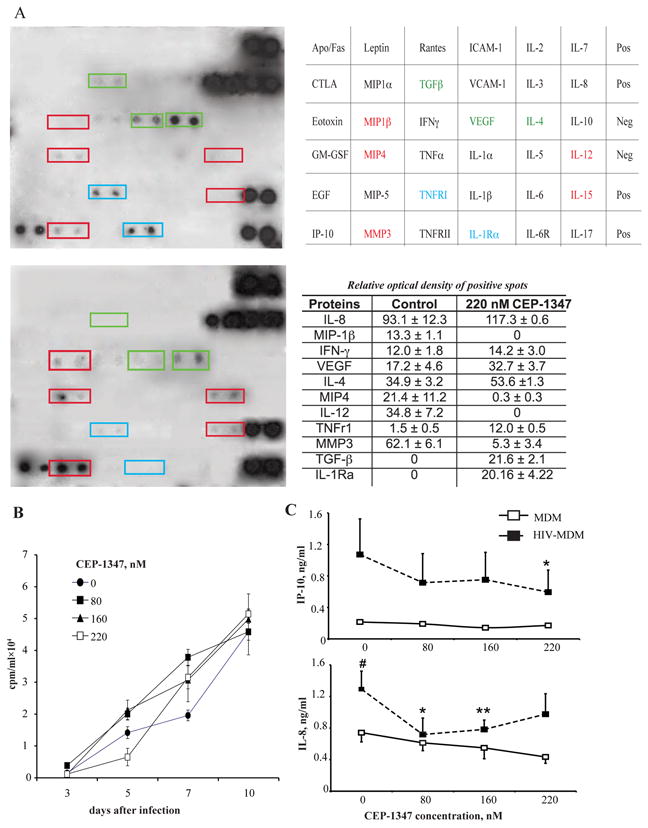
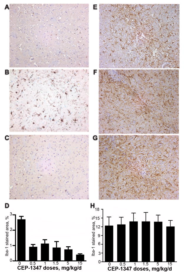
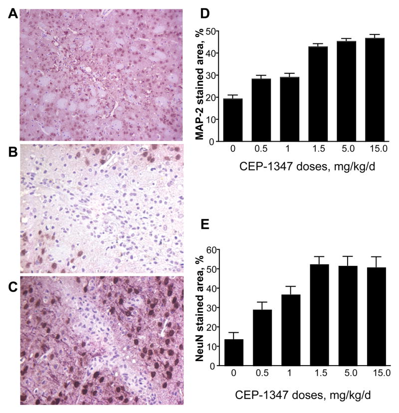
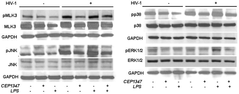
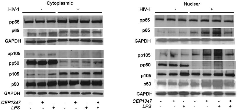
When the nervous system is infected with HIV-1, it commonly results in neuroinflammation leading to overt neuronal dysfunction and subsequent cognitive and behavioral impairments. The multifaceted disease process, now referred to as HIV-1-associated neurocognitive disorders (HAND), provides a range of molecular targets for adjunctive therapies. One is CEP-1347, an inhibitor of mixed lineage kinases that elicits neuroprotective and anti-inflammatory responses in models of neurodegenerative diseases. Since HAND is associated with inflammatory encephalopathy induced by virus infection and mononuclear phagocytes (perivascular macrophages and microglia) immune activation, we investigated whether CEP-1347 could ameliorate disease in laboratory models of HAND. We now demonstrate that CEP-1347 reduces the levels of secreted proinflammatory cytokines and chemokines in HIV-1-infected human macrophages and attenuates dose-dependent neurotoxicity in rodent cortical neurons. CEP-1347-treated mice readily achieve therapeutic drug levels in peripheral blood. HIV-1 encephalitis (HIVE) mice, where human virus-infected monocyte-derived macrophages are stereotactically injected into the basal ganglia of CB17 severe combined immunodeficient mice, received daily intraperitoneal injections of CEP-1347. Here, CEP-1347 treatment of HIVE mice showed a dose-dependent reduction in microgliosis. Dendritic integrity and neuronal loss were sustained and prevented, respectively. These results demonstrate that CEP-1347 elicits anti-inflammatory and neuroprotective responses in an HIVE model of human disease and as such warrants further study as an adjunctive therapy for human disease.


Figures

Figure 1
Figure 1
CEP-1347 and MDM secretory and viral replication activities. Effect of CEP-1347 on HIV-1 replication, growth factor, chemokine, pro-inflammatory cytokine and enzyme secretion by human monocyte derived macrophages (MDM). A, TransSignal human cytokine antibody array 3.0 by Panomics and results table. At 5-day post-HIV-1ADA infection, human MDM were treated with 220 nM CEP-1347 (upper blot) or vehicle (lower blot) for 45 min. Cells were washed to remove CEP-1347, and supernatants were harvested 24 hr later. Each cytokine on array is composed of 2 horizontal dots. Data is representative of 3 independent experiments (mean ± SEM). B, CEP-1347 was administered to cultures at a concentration of 0 – 220 nM 24 hrs before, at the time of, and 4 hrs after HIV-1ADA infection. Monocytes were cultivated for seven days, then infected at a MOI of 0.01. Supernatant fluids were collected and assayed for RT activities from zero to 10 days after viral infection. Values represent counts per minute (mean ± SEM) of samples from six independent cultures and are representative of three independent separate experiments when CEP-1347 was added simultaneously with infection. The same results were obtained with drug administration before and after infection. C, In addition, Cytometric Bead Array showed down-regulation of IL-8 and CXCL10/IP-10. Values represent concentrations of chemokines in supernatants collected from three independent experiments, when CEP-1347 was added simultaneously with infection (mean ± SEM). # - P < 0.05 difference between uninfected and HIV-1-infected MDM. *P < 0.05, differences between untreated and treated infected cells. **P < 0.01, differences between untreated and treated infected cells.
Figure 2
Figure 2
CEP-1347 neuroprotective activities. Human MDMs were infected with HIV-1ADA (MOI of 0.01) and treated simultaneously with CEP-1347 (80 or 160 nM). Conditioned media was collected at day 5 after infection. Twenty four hrs before conditioned media harvest MDM were washed to remove CEP-1347 and neurobasal media was added to the cultures. Conditioned media thus collected were added to primary MCN to measure neurotoxicity levels. Neurons were stained immunocytochemically for MAP-2 (red) and NeuN (green) to assess neuronal integrity. A, Primary MCN culture treated with conditioned media from control MDM. B, Primary MCN cultures cultivated with HIV-1 infected MDM conditioned media. These neuronal cultures demonstrate both decreased dendritic density (MAP-2 immunostaining) and reduced neural density (number of NeuN positive cells). C, HIV-1 infected MDM conditioned media that received 80 nM CEP-1347 show partial protection of dendritic and neural density. D, Conditioned media from HIV-1 infected MDM treated with 160 nM CEP-1347 show protection of dendritic and neural densities. Original magnification x400.
Figure 3
Figure 3
Quantitative analyses of CEP-1347 induced neuroprotective activities. Quantitative analyses of MAP-2 and NeuN staining in primary MCN culture treated with conditioned media from CEP-1347 treated, HIV-1-infected MDM. A, Analysis of dendritic density, as assessed by MAP-2 staining and neural density, measured by NeuN staining. Data demonstrate MFI at 617 nm (MAP-2) and 519 nm (NeuN) for culture wells containing treated MCN. Values are mean ± SEM for quadruplicate cultures. Data are representative of three independent experiments using three different donors. B, LDH concentration from replicate primary cortical neural cultures with CM from CEP-1347 treated, HIV-1-infected MDM; values are mean ± SEM of quadruplicate cultures. Data expressed are percentage changes in LDH release compared to MCN treated with 1nM St, 100%. Data are representative of three independent experiments using three different donor conditioned medias.
Figure 4
Figure 4
Pharmacokinetic analyses of CEP-1347 levels in plasma of experimentally treated mice. The plasma concentration of CEP-1347 was measured following i.p. dosing of mice with 1.5 or 15.0 mg/kg of CEP-1347. For this experiment, both C57Bl/6 and CB17 SCID mice were treated. However, the CB17 SCID mice were treated only with the lower 1.5 mg/kg dose of CEP-1347. Blood samples were collected at selected time points (0.5, 1, 2, 4, 6 and 8 h), and plasma was separated for analyses. Samples were analyzed by liquid chromatography/mass spectrometry. Values are mean ± SEM from 4 animals. Data are represented as a line graph (n = 4 mice/treatment).
Figure 5
Figure 5
CEP-1347 suppresses neuroinflammatory responses in murine HIVE. SCID mice were injected with HIV-1ADA infected MDM with/out CEP-1347 treatment. MDM were propagated in suspension cultures and infected with HIV-1ADA at an MOI of 0.1 for 5 days prior to brain injections. All animals were sacrificed seven days after i.c. cell injection. Immunohistological images of the basal ganglia are shown. Serial 5-μM sections containing the injection area were stained for Iba-1 (A, B, C) and GFAP (E, F, G). A, Immunohistological stain for Iba-1 in sham-operated murine brain. B, Replicate image to (A) of area adjacent to injection site in HIVE murine brain stained for Iba-1; note the increased in Iba-1 reactive microglia. C, Immunohistological stain for Iba-1 in tissue adjacent to the injection site from HIVE murine brain treated with 15 mg/kg/d CEP-1347; note the reduction in Iba-1 staining compared to B. D, Quantitation of Iba-1 staining. Values are mean ± SEM obtained from analyzing four brain tissue sections for each experimental animal (4 mice/group). E, Immunohistological image of uninfected murine brain stained for GFAP. F, Untreated HIVE brain and G, HIVE brain treated with 15 mg/kg/d CEP-1347. H, Quantification of GFAP staining. Values are mean ± SEM obtained from analyzing three brain tissue sections in each experimental animal (4 mice/group).
Figure 6
Figure 6
CEP-1347 elicits a neuroprotective effect in HIVE mice. Tissue sections from HIVE mice were stained for MAP-2 (pink) and NeuN (brown) (A, B, C). Neuronal integrity was assessed in areas of focal encephalitis in CEP-1347 treated mice. A, Immunohistochemical stain for MAP-2 and NeuN in control sham-operated mice. B, Immunohistological image of HIV-1-infected murine brain stained for MAP-2 and NeuN, 7 days post injection. C, Immunohistological image of the brain from an HIV-1-infected mouse treated with 15 mg/kg/d of CEP-1347, stained for MAP-2 and NeuN, 7 days post injection. Original magnification 200×. D, Quantitation of MAP-2 immunoreactivity with increasing doses of CEP-1347. Values are mean ± SEM from multiple images obtained at original magnification 200× per mouse (4 mice/group). E, Quantitation of NeuN immunoreactivity with increasing doses of CEP-1347. Values are mean ± SEM from multiple images per mouse (4 mice/group).
Figure 7
Figure 7
MAPK kinase pathways in HIV-1ADA infected human MDM treated with CEP-1347. Human monocytes collected after centrifugal elutriation were cultured for seven days in 1,000 U/ml MCSF then infected with HIV-1ADA viral stock at an MOI of 0.01 for 6 h with/out 220 nM CEP-1347. CEP-1347 was continued through the experiment. Uninfected MDM with or without CEP-1347 was used as controls. Viral replication continued for 5 days. Viral infection at 5 days showed RT activity 10-fold above background levels. Replicate control and HIV-1 infected MDM were treated with LPS at 100 ng/ml for 24 h when all cells were harvested for Western blot assays. Cell lysates were analyzed using Abs specific MLK3, phospho-MLK3, ERK1/2, phospho-ERK1/2, p38, phospho-p38, JNK and phospho-JNK. The data presented here is a representative of four independent experiments.
Figure 8
Figure 8
CEP-1347 does not affect NF-κB pathways in HIV-1 infected human MDM. Human monocytes collected after centrifugal elutriation were cultured for seven days in 1,000 U/ml MCSF then infected with HIV-1ADA viral stock at an MOI of 0.01 for 6 h with or without CEP-g1347 at 220 nM. CEP-1347 was continued through the experiment. Uninfected MDM with or without CEP-1347 was used as controls. Viral infection at 5 days showed RT activity 10-fold above background levels. Replicate control and HIV-1 infected MDM were treated with LPS at 100 ng/ml for 24 h when all cells were harvested for Western blot assays. Cells were fractionated into cytosolic and nuclear fractions using Nuclear/Cytosol fractionation Kit (BioVision, Cat. No- K266-25). The fractionation was performed according to the manufacturers instructions. Both cytosolic and nuclear fractions were then analyzed by using specific Ab to p65, p105/p50 and its phophorylated derivatives (Cell Signaling Technology). For all analyses GAPDH was used as loading control. The data presented here is a representative of three independent experiments.

Similar articles

Cited by

References

    1. Lipton SA, Gendelman HE. Dementia associated with the acquired immunodeficiency syndrome. New Engl J Med. 1995;16:934–940. - PubMed
    1. Michaels J, Sharer LR, Epstein LG. Human immunodeficiency virus type 1 (HIV-1) infection of the nervous system: a review. Immunodefic Rev. 1988;1:71–104. - PubMed
    1. Everall IP, Luthert PJ, Lantos PL. Neuronal loss in the frontal cortex in HIV infection [see comments] Lancet. 1991;337:1119–1121. - PubMed
    1. Swindells S, Zheng J, Gendelman HE. HIV-associated dementia: new insights into disease pathogenesis and therapeutic interventions. Aids Patient Care STDS. 1999;13:153–163. - PubMed
    1. Kure K, Llena JF, Lyman WD, Soeiro R, Weidenheim KM, Hirano A, Dickson DW. Human immunodeficiency virus-1 infection of the nervous system: an autopsy study of 268 adult, pediatric, and fetal brains. Hum Pathol. 1991;22:700–710. - PubMed

Publication types

MeSH terms