. 2009 Dec 22;106(51):21942-7.
doi: 10.1073/pnas.906637106. Epub 2009 Dec 4.

Sleep-wake regulation and hypocretin-melatonin interaction in zebrafish

Affiliations

Sleep-wake regulation and hypocretin-melatonin interaction in zebrafish

Lior Appelbaum et al. Proc Natl Acad Sci U S A. .

Abstract

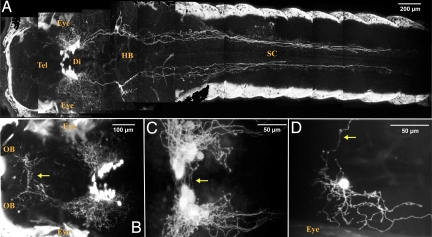
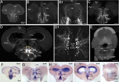
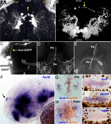
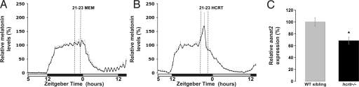
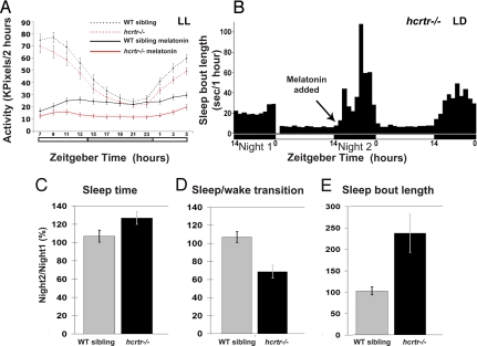
In mammals, hypocretin/orexin (HCRT) neuropeptides are important sleep-wake regulators and HCRT deficiency causes narcolepsy. In addition to fragmented wakefulness, narcoleptic mammals also display sleep fragmentation, a less understood phenotype recapitulated in the zebrafish HCRT receptor mutant (hcrtr-/-). We therefore used zebrafish to study the potential mediators of HCRT-mediated sleep consolidation. Similar to mammals, zebrafish HCRT neurons express vesicular glutamate transporters indicating conservation of the excitatory phenotype. Visualization of the entire HCRT circuit in zebrafish stably expressing hcrt:EGFP revealed parallels with established mammalian HCRT neuroanatomy, including projections to the pineal gland, where hcrtr mRNA is expressed. As pineal-produced melatonin is a major sleep-inducing hormone in zebrafish, we further studied how the HCRT and melatonin systems interact functionally. mRNA level of arylalkylamine-N-acetyltransferase (AANAT2), a key enzyme of melatonin synthesis, is reduced in hcrtr-/- pineal gland during the night. Moreover, HCRT perfusion of cultured zebrafish pineal glands induces melatonin release. Together these data indicate that HCRT can modulate melatonin production at night. Furthermore, hcrtr-/- fish are hypersensitive to melatonin, but not other hypnotic compounds. Subthreshold doses of melatonin increased the amount of sleep and consolidated sleep in hcrtr-/- fish, but not in the wild-type siblings. These results demonstrate the existence of a functional HCRT neurons-pineal gland circuit able to modulate melatonin production and sleep consolidation.


Conflict of interest statement

The authors declare no conflict of interest.

Figures

Fig. 1.
Fig. 1.
Zebrafish HCRT neurons are glutamatergic. (A–D) and close-ups (A1–D1) Double fluorescent ISH between hcrt mRNA and fast neurotransmitter phenotype markers as visualized using confocal microscopy on adult brain sections (reconstructed stacks of 0.5- or 1-μm sections). (C2–C4) and (D2–D4): single-plane, high-magnification pictures of hcrt cells (green, C2, D2), vglut2a or -b cells (red, C3, D3), and merged views (C4, D4). Arrowheads indicate cells coexpressing hcrt and vglut2a or vglut2b. Note the frequent colocalization. Absence of coexpression is occasionally observed (arrow). [Scale bar, 100 μm (A–D), 20 μm (A1–D1, C2–C4, and D2–D4).]
Fig. 2.
Fig. 2.
A stable hcrt:EGFP transgenic line reveals the entire HCRT circuit of a living larva. (A–C) Two-photon imaging of a 7dpf stable transgenic larva with head to the left, dorsal views. (A and close-up B) Composite picture showing the HCRT cell bodies in the diencephalon (Di) and their processes in the hindbrain (HB) and spinal cord (SC) and in the telencephalon (Tel) directed toward the olfactory bulbs (OB). In A and B, white areas on both sides of the larva correspond to skin autofluorescence. Commissural projections are observed ventrally to the HCRT cell body clusters (C, arrow) and in the anterior telencephalon (B, arrow, commissura anterior). (D) Mosaic expression of a nonintegrated hcrt:EGFP transgene allowing the observation of a single HCRT neuron harboring both commissural (arrow) and ipsilateral processes.
Fig. 3.
Fig. 3.
Adult zebrafish HCRT circuit. (A–E) Confocal imaging of 100 μm transversal brain sections from a stable hcrt:EGFP transgenic adult fish (reconstructed stacks of 0.5- or 1-μm sections). Note the compact organization of the HCRT cell bodies in the periventricular hypothalamus (D and dashed-box close up in D1). (F–J) hcrtr mRNA ISH pattern in equivalent brain sections to A–E. Note the similar distribution of HCRT fibers and hcrtr mRNA. Compare sections of telencephalon (A vs. F), telencephalon-midbrain boundary (B and B1 vs. G), anterior diencephalon and mesencephalon (C vs. H), mid diencephalon and mesencephalon (D vs. I), rhombencephalon (E vs. J). Chab, commissura habenularum; Ctec, commissura tecti; Cpost, commissura posterior; CC, crista cerebellaris; CP, central posterior thalamic nucleus; D, dorsal telencephalic area; Dm, medial zone of D; Dc, central zone of D; Dl, lateral zone of D; DIL, diffuse nucleus of the inferior lobe; Ha, habenula; Hv, ventral zone of the periventricular hypothalamus; Hd, dorsal zone of the periventricular hypothalamus; IMRF, intermediate reticular formation; PG, preglomerular nucleus; PGZ, periventricular gray zone of the optic tectum; PPp, parvocellular preoptic nycleus, posterior part; PS, pineal stalk; SO, secondary octaval population; TBS, tractus bulbospinalis; TeO, optic tectum; TPp, periventricular nucleus of posterior tuberculum; V, ventral telencephalic area; Vv, ventral nucleus of V; Vd, dorsal nucleus of V.
Fig. 4.
Fig. 4.
The HCRT-pineal gland circuit. (A and B) Dorsal and frontal views of the brain of a 7 dpf hcrt:EGFP transgenic larva imaged by two-photon microscopy. HCRT axons (arrows) projecting toward the pineal gland are observed. (C) A dorsal image of 6 dpf transgenic larva carrying two transgenes; an EGFP reporter driven by hcrt (hcrt:EGFP) and the pineal-specific aanat2 (aanat2:EGFP) promoters, demonstrate direct axon projection (arrow) to the pineal gland. (D and E) Close-ups of two adjacent transversal hcrt:EGFP adult brain sections showing HCRT projections to the habenula and the pineal gland stalk. (F) Lateral and (G and H) dorsal views of whole-mount in situ hybridization of 2-dpf embryos. (F) hcrtr mRNA is expressed in several regions of the brain (16) including the pineal gland (arrow). Double ISH experiment with aanat2 demonstrates that hcrtr is expressed in the pineal gland during the day (G) and the night (H). Similarly, in adult animals, hcrtr is expressed in the pineal gland (I). aanat2 (J) and egfp (K) probes were used as positive and negative controls, respectively. Adult pineal glands (I–K) were removed with the upper skull and skin hence presence of brown melanophores cells in the preparations.
Fig. 5.
Fig. 5.
HCRT modulates pineal gland melatonin production. (A and B) HCRT induces melatonin release from cultured zebrafish pineal glands. (A) Normal circadian rhythm of melatonin release of melatonin from zebrafish pineal glands cultured in constant darkness. MEM medium application does not affect melatonin production in 12 control pineal glands. (B) zebrafish HCRT-1 application (10−6 M) stimulates melatonin production (ZT 21–23) (n = 8, P < 0.05). The production lasts for the duration of HCRT application. (C) Quantitative PCR analysis of aanat2 mRNA level in six wild-type sibling and 6 hcrtr−/− pineal glands collected during the night (ZT 16). Note the significant decrease (31%, P < 0.05) of aanat2 expression in pineal glands devoid of functional HCRT input.
Fig. 6.
Fig. 6.
hcrtr−/− fish are hypersensitive to the melatonin sleep-promoting effect. (A) Larvae were kept under constant dim light conditions (LL, <10lux) in the monitoring system (white bars represent the subjective day period). hcrtr−/− and wild-type sibling 5 dpf larvae demonstrate similar rhythmic activity that peak during the day. hcrtr−/− larvae were significantly more sensitive to melatonin's (1 μM) hypnotic effect. (B–E) Adult fish were kept under LD cycle (represented by white and black bars) in AFSRS [SI Materials and Methods and (16)] and fine sleep architecture was analyzed. (B) Representative sleep bout pattern of an adult hcrtr−/− mutant. Subthreshold dose of melatonin (1 μM) was added at the beginning of the second night (arrow) and sleep parameters were compared to first night. hcrtr−/− adults were more sensitive to melatonin as, after its administration, total sleep time increased (C), the number of sleep/wake transition decreased (D) and sleep bout length increased (E), as compared to wild-type siblings.

References

    1. de Lecea L, et al. The hypocretins: Hypothalamus-specific peptides with neuroexcitatory activity. Proc Natl Acad Sci USA. 1998;95:322–327. - PMC - PubMed
    1. Sakurai T, et al. Orexins and orexin receptors: A family of hypothalamic neuropeptides and G protein-coupled receptors that regulate feeding behavior. Cell. 1998;92:573–585. - PubMed
    1. Peyron C, et al. Neurons containing hypocretin (orexin) project to multiple neuronal systems. J Neurosci. 1998;18:9996–10015. - PMC - PubMed
    1. de Lecea L, Sutcliffe JG. The hypocretins and sleep. FEBS J. 2005;272:5675–5688. - PubMed
    1. van den Pol AN. Hypothalamic hypocretin (orexin): Robust innervation of the spinal cord. J Neurosci. 1999;19:3171–3182. - PMC - PubMed

Publication types

MeSH terms