. 2010 Mar;38(4):1135-48.
doi: 10.1093/nar/gkp1097. Epub 2009 Dec 4.

Opposing regulatory roles of phosphorylation and acetylation in DNA mispair processing by thymine DNA glycosylase

Affiliations

Opposing regulatory roles of phosphorylation and acetylation in DNA mispair processing by thymine DNA glycosylase

Ryan D Mohan et al. Nucleic Acids Res. 2010 Mar.

Abstract

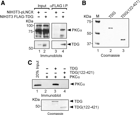
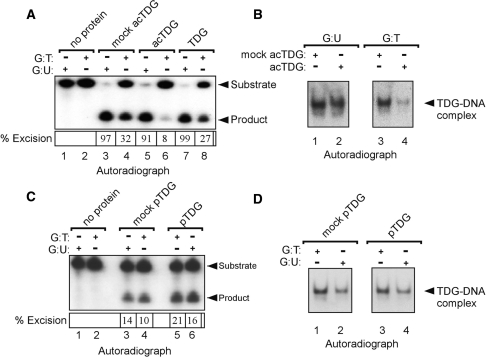
CpG dinucleotides are mutational hotspots associated with cancer and genetic diseases. Thymine DNA glycosylase (TDG) plays an integral role in CpG maintenance by excising mispaired thymine and uracil in a CpG context and also participates in transcriptional regulation via gene-specific CpG demethylation and functional interactions with the transcription machinery. Here, we report that protein kinase C alpha (PKCalpha) interacts with TDG and phosphorylates amino-terminal serine residues adjacent to lysines acetylated by CREB-binding protein (CBP) and p300 (CBP/p300). We establish that acetylation and phosphorylation are mutually exclusive, and their interplay dramatically alters the DNA mispair-processing functions of TDG. Remarkably, acetylation of the amino-terminal region abrogates high-affinity DNA binding and selectively prevents processing of G:T mispairs. In contrast, phosphorylation does not markedly alter DNA interactions, but may preserve G:T processing in vivo by preventing CBP-mediated acetylation. Mutational analysis suggests that the acetyl-acceptor lysines are not directly involved in contacting DNA, but may constitute a conformationally sensitive interface that modulates DNA interactions. These findings reveal opposing roles of CBP/p300 and PKCalpha in regulating the DNA repair functions of TDG and suggest that the interplay of these modifications in vivo may be critically important in the maintenance of CpG dinucleotides and epigenetic regulation.


Figures

Figure 1.
Figure 1.
Phorbol ester-stimulated phosphorylation of TDG in living cells. (A) Illustration of the functional domains of mouse TDG and sites of posttranslational modification. The central-conserved glycosylase domain is sufficient for processing of G:U mispairs while a more divergent amino-terminal extension is required for tight DNA binding and G:T processing (20,21). Two SUMO-binding motifs (SBM1 and SBM2) and the sumoylation site (K341) are shown. A lysine-rich regulatory region located in the amino-terminus is acetylated by CBP/p300 at four distinct lysines (K70, K94, K95 and K98) (15). Putative protein kinase C (PKC) α/β/γ-phosphorylation sites (consensus [S/T-X-[R/K]) within a sequence (boxed) conserved in mouse, rat and human TDG are indicated by asterisks. Complete sequence alignments and accession numbers are found in Figure S1. (B) 2D PAGE analysis of cellular TDG demonstrates PMA-dependent alterations in apparent molecular weight and isoelectric point (pI). Cell lysates were prepared from NIH3T3 cells stimulated with PMA and then separated by 2D PAGE. TDG was detected by immunoblotting with a TDG-specific antibody. (B) In vivo metabolic labelling of transiently expressed TDG with 32P-orthophosphate. Transfected NIH3T3 fibroblasts were grown in serum-free media and metabolically labelled with or without PMA treatment. One population of transfected cells was pretreated with a PKCα/β inhibitor (Gö6976) before PMA stimulation. Immunoprecipitated TDG was fractionated by SDS–PAGE and analysed by phosphorimaging (upper panel) and immunoblotting (lower panel).
Figure 2.
Figure 2.
Subcellular localization of TDG and PKCα in NIH3T3 fibroblast and P19 EC cells. (A) Subcellular localization of endogenous TDG and PKCα in NIH3T3 cells. Serum-starved cells were treated with PMA or DMSO and subsequently immunostained for TDG and PKCα. (B) Undifferentiated P19 embryonic carcinoma cells (not treated) were immunostained to detect endogenous TDG and PKCα. Representative optical sections generated by epifluorescence microscopy are shown. The fluorescence intensity plot illustrates the coincidence of peak fluorescence for TDG (CY3, red) and PKCα (FITC, green).
Figure 3.
Figure 3.
PKCα associates directly with TDG. (A) PKCα coimmunoprecipitates with stably expressed TDG in NIH3T3 fibroblasts. Immunoprecipitations using anti-FLAG affinity resin were carried out on whole-cell extracts prepared from PMA-treated cells stably expressing FLAG–TDG or control cells transduced with the empty expression vector. Aliquots of the cell lysates and immunoprecipitated proteins were immunoblotted for PKCα and TDG. (B) Coomassie staining of polyhistidine-tagged TDG and TDG(122–421) used for in vitro protein interaction studies. (C) In vitro association of recombinant TDG and PKCα requires amino-terminal residues 1–121 of TDG. Approximately 1 µg polyhistidine-tagged TDG or TDG(121–421) were incubated with 10 ng of PKCα and subjected to pull down with nickel-affinity resin. As a control, PKCα was also incubated with beads alone. Bound proteins were subjected to SDS–PAGE and immunoblotting with a PKCα-specific antibody (upper panel). Binding of poly-histidine-tagged TDG and TDG(122–421) to the nickel-affinity beads was confirmed by Coomassie staining (lower panel).
Figure 4.
Figure 4.
PKCα phosphorylates TDG on serines 96 and 99. (A) PKCα-mediated phosphorylation of TDG requires the amino-terminal region. In vitro phosphorylation reactions were performed in the presence of γ32P-ATP using 2 µg of TDG (lane 1) or TDG(122–421) (lane 2) and 0.3 ng of recombinant PKCα. Reaction products were fractionated by SDS–PAGE and incorporation of 32P was detected by phosphorimaging. (B) Delineation of the phosphorylated region using peptide probes. Equimolar amounts of TDG peptides (residues 68–91 and 91–107) and PKCα peptide substrate from glycogen synthetase (residues 1–8 and designated GS 1–8) along with the FLAG peptide were reacted with PKCα and analysed as indicated earlier. (C) Identification of phosphoacceptor residues by alanine substitution. In vitro phosphorylation of the TDG(91–107) (1 µg) peptide and alanine-substituted derivatives was performed and analysed by SDS–PAGE and phosphorimaging (top panel). Quantification of signal intensity is displayed in the bottom panel. Lysines acetylated by CBP/p300 are indicated with asterisks. (D) Dual alanine (lane 2) or aspartate (lane 3) substitutions of serine 96 and 99 reduces PKCα-mediated phosphorylation in full-length TDG. Recombinant TDG and the indicated substitution mutants (2 µg) were phosphorylated in vitro and analysed by SDS–PAGE and autoradiography. (E) In vivo metabolic labelling of transiently expressed TDG and S96-99D mutant with 32P-orthophosphate. Transfected NIH3T3 fibroblasts grown in serum-free media were metabolically labelled for 2.5 h, which includes treatment with either vehicle or PMA during the final 30 min of labelling. Proteins were immunoprecipitated with anti-FLAG resin and then fractionated by SDS–PAGE and phosphorimaged (upper panel). Aliquots of the immunoprecipitates were analysed by immunobloting to ensure equal loading of TDG (lower panel).
Figure 5.
Figure 5.
Acetylation and phosphorylation are mutually exclusive. (A) Acetylated TDG (acTDG) is refractory to phosphorylation by PKCα. Recombinant polyhistidine-tagged TDG was acetylated in vitro with CBP and then purified by nickel-affinity chromatography. acTDG was quantified by immunoblotting and ∼100 ng was used in phosphorylation reactions that included γ32P-ATP. Reaction products were analysed by SDS–PAGE and phosphorimaging. (B) Phosphorylated TDG (pTDG) is refractory to acetylation by CBP. pTDG was purified as indicated above and reacted with CBP and in the presence of 14C-acetyl coenzyme A (AcCoA). (C) Reduced in vitro phosphorylation of the K94-95-98A mutant. (D) Reduced in vitro acetylation of the S96-99D mutant. Recombinant proteins (1 µg) were phosphorylated or acetylated in vitro as described above. (E) Inhibition of in vivo TDG phosphorylation by coexpression of CBP or substitution of positively charged acetyl-acceptor lysines. HEK 293T cells were transfected with the indicated expression vector and metabolically labelled with and without stimulation with PMA.
Figure 6.
Figure 6.
DNA binding prevents CBP-mediated acetylation of TDG. (A) Dose-dependent inhibition of TDG acetylation by duplex oligonucleotides. TDG (150 ng) was preincubated with the indicated duplex oligonucleotides for 30 min on ice and then acetylated in vitro with CBP (100 ng) and 14C-AcCoA. Reaction products were analysed by SDS–PAGE and autoradiography. (B) DNA-dependent inhibition of CBP-mediated acetylation is specific to TDG. In vitro acetylation was performed with TDG (150 ng), GST-p53 (150 ng) and SET/TAF-1β (150 ng) recombinant proteins in the presence or absence of 200 ng of G:T mispaired oligonucleotide. (C) Phosphorylation of TDG in the presence of duplex oligonucleotides. In vitro phosphorylation reactions were performed as described in Figure 4 using TDG preincubated with 200 ng of the indicated oligonucleotides. (D) Alanine substitution of acetyl acceptor lysines enhances DNA binding. Electrophoretic mobility shift assay was carried out with radiolabelled duplex oligonucleotides bearing either a G:U or a G:T mispair with 25 ng of recombinant wild-type TDG or TDG(K94-95-98A).
Figure 7.
Figure 7.
Divergent effects of acetylation and phosphorylation on DNA mispair processing. (A) Acetylation of TDG selectively abrogates G:T processing. Purified acetylated TDG (acTDG) (25 ng) was prepared as described in Figure 5, and base-excision assays were carried out using asymmetrically radiolabelled duplex oligonucleotides (10 ng) bearing either G:U or G:T mispairs. Reaction products were treated with alkali to cleave the abasic sites and analysed by denaturing PAGE and autoradiography. (B) acTDG does not stably bind oligonucleotides bearing a G:T mispair. Aliquots of base-excision reactions described above were subjected to electrophoretic mobility-shift assays to determine binding to G:U or G:T mispaired oligonucleotides. (C) Phosphorylation of TDG does not detectably alter G:T/U processing activity. Phosphorylated (pTDG) and mock pTDG (12 ng) were tested for ability to excise mispaired uracil and thymine. (D) DNA-binding analysis of phosphorylated and mock pTDG. Aliquots of the base-excision reactions were subjected to electrophoretic mobility-shift analysis. Supplementary Figures S4 and S5 show images of EMSA gels that include unbound DNA probe.
Figure 8.
Figure 8.
Cross-talk between TDG posttranslational modifications. Previous studies have shown that sumoylation of human and mouse TDG induces a dramatic increase in G:U processing activity by promoting enzyme turnover (19,20). In contrast, sumoylation (19,20) or acetylation by CBP (this study) abrogate DNA binding and G:T processing (19,20). TDG sumoylation also drastically reduces interactions with CBP/p300, thereby preventing efficient acetylation (20). The present studies reveal that the phosphorylation of serine residues adjacent to acetyl-acceptor lysines by PKCα prevents acetylation by CBP and may preserve G:T processing in vivo.

Similar articles

Cited by

References

    1. Klose RJ, Bird AP. Genomic DNA methylation: the mark and its mediators. Trends Biochem. Sci. 2006;31:89–97. - PubMed
    1. Pfeifer GP. Mutagenesis at methylated CpG sequences. Curr. Top Microbiol. Immunol. 2006;301:259–281. - PubMed
    1. Lindahl T. Instability and decay of the primary structure of DNA. Nature. 1993;362:709–715. - PubMed
    1. Pfeifer GP. p53 mutational spectra and the role of methylated CpG sequences. Mutat. Res. 2000;450:155–166. - PubMed
    1. Petitjean A, Mathe E, Kato S, Ishioka C, Tavtigian SV, Hainaut P, Olivier M. Impact of mutant p53 functional properties on TP53 mutation patterns and tumor phenotype: lessons from recent developments in the IARC TP53 database. Hum. Mutat. 2007;28:622–629. - PubMed

Publication types

Substances