. 2010 Jan 5;107(1):332-7.
doi: 10.1073/pnas.0912730107. Epub 2009 Dec 4.

TRPM1 is a component of the retinal ON bipolar cell transduction channel in the mGluR6 cascade

Affiliations

TRPM1 is a component of the retinal ON bipolar cell transduction channel in the mGluR6 cascade

Chieko Koike et al. Proc Natl Acad Sci U S A. .

Abstract

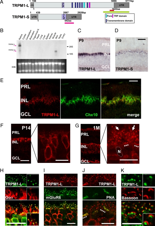
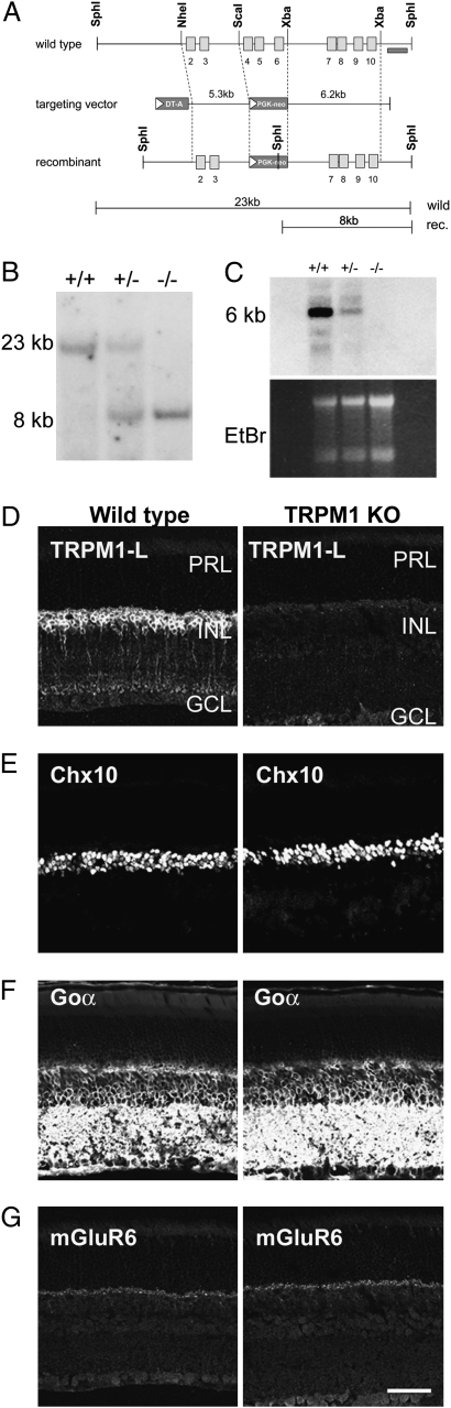
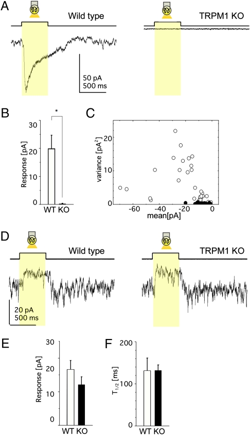
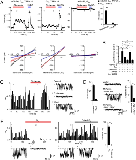
An essential step in intricate visual processing is the segregation of visual signals into ON and OFF pathways by retinal bipolar cells (BCs). Glutamate released from photoreceptors modulates the photoresponse of ON BCs via metabotropic glutamate receptor 6 (mGluR6) and G protein (Go) that regulates a cation channel. However, the cation channel has not yet been unequivocally identified. Here, we report a mouse TRPM1 long form (TRPM1-L) as the cation channel. We found that TRPM1-L localization is developmentally restricted to the dendritic tips of ON BCs in colocalization with mGluR6. TRPM1 null mutant mice completely lose the photoresponse of ON BCs but not that of OFF BCs. In the TRPM1-L-expressing cells, TRPM1-L functions as a constitutively active nonselective cation channel and its activity is negatively regulated by Go in the mGluR6 cascade. These results demonstrate that TRPM1-L is a component of the ON BC transduction channel downstream of mGluR6 in ON BCs.


Conflict of interest statement

The authors declare no conflict of interest.

Figures

Fig. 1.
Fig. 1.
The molecular analysis and expression of mouse TRPM1-L. (A) Schematic diagrams of full-length mouse TRPM1-L and TRPM1-S genes and their ORFs. Light gray boxes represent ORF, and light purple boxes indicate exon 14 (L and S). Green and pink bars indicate sequences used for TRPM1-L- or TRPM1-S-specific probes, respectively. The red bar indicates amino acid sequence used for generating anti-TRPM1-L antibody. (B) Northern blot analysis of mouse TRPM1 transcription in adult mouse tissues. The sizes of TRPM1-L and TRPM1-S transcripts are ≈6 kb and 3 kb, respectively. Both TRPM1-L and -S transcripts were detected in the retina; however, only the TRPM1-S transcript was detected in the skin. (Lower) Ethidium bromide staining of RNA. Each lane contains ≈10 μg of total RNA. Trigeminal N., trigeminal nucleus. (C and D) ISH analysis of mouse TRPM1 in the postnatal retina. TRPM1-L-specific signal was detected in the INL at P9 (C). TRPM1-S was detected in the INL at P9 (D). PRL, photoreceptor layer; INL, inner nuclear layer; GCL, ganglion cell layer. (Scale bar: 100 μm.) (E) ISH of TRPM1-L mRNA and following immunostaining of anti-Chx10 antibody, a pan-BC marker. (Scale bar: 50 μm.) (F and G) Immunostaining with an antibody against TRPM1-L exhibited TRPM1-L signals in cell bodies of retinal BCs at P14 (F) and at dendritic tips of retinal BCs (stars and arrows) at 1 M (G). N, nucleus of a BC. (Scale bars: 50 μm.) (HK) Confocal images of OPLs double-labeled with anti-TRPM1-L antibody and other retinal markers. TRPM1-L-positive puncta were localized at the tips of Goα distribution (H). TRPM1-L-positive puncta were colocalized with mGluR6 staining (I). Continuous puncta marked with TRPM1-L were colocalized with PNA (stars) (J). TRPM1-L-positive puncta were surrounded by synaptic ribbons stained with bassoon (K). (Scale bars: 10 μm.)
Fig. 2.
Fig. 2.
Generation of TRPM1−/− mouse by targeted gene disruption. (A) Strategy for the targeted deletion of TRPM1 gene. The open boxes indicate exons. Exons 4–6 were replaced with the PGK-neo cassette. The probe used for Southern blot analysis is shown as a dark bar. (B) Southern blot analysis of genomic DNA. SphI-digested genomic DNA was hybridized with a 3′ outside probe, detecting 23-kb WT and 8-kb mutant bands. (C) Northern blot analysis of total RNA extracted from the retina derived from WT, TRPM1+/−, and TRPM1−/− mice using cDNA probe derived from exons 3–9. (DG) Immunostaining of WT and TRPM1−/− retinal sections at 1 M. The TRPM1-L signal in the INL of WT mouse retina disappeared in the TRPM1−/− mouse retina (D). Immunohistochemical analysis using antibodies to Chx10 [pan-BC nuclei marker (E)], Goα [ON BC dendrite marker (F)], and mGluR6 [ON BC dendrite tip marker (G)] showed no obvious difference between WT and TRPM1−/− mice retinas. PRL, photoreceptor layer; INL, inner nuclear layer; GCL, ganglion cell layer. (Scale bar: 50 μm.)
Fig. 3.
Fig. 3.
TRPM1-L is essential for ON BC photoresponses. (AC) Data from ON BCs in the retinal slice preparation. (A) Photoresponses of ON BCs from WT (Left) and TRPM1−/− (Right) mice (holding potential at –62 mV). Each trace illustrates the average of three responses. (B) Mean ± SEM of photoresponses from WT (15 cells, 12 mice) and TRPM1−/− (9 cells, 8 mice) mice. *, P < 0.05. (C) The variance against the mean (the leak current not subtracted) of the dark membrane current fluctuations obtained from WT (open circles: 34 traces, 15 cells) and TRPM1−/− (filled circles: 26 traces, 9 cells) mice. (DF) Data from OFF BCs in the retinal slice preparation. (D) Photoresponses of OFF BCs from WT (Left) and TRPM1−/− (Right) mice under similar recording conditions as in A. Each trace illustrates the average of two responses. No significant difference in the response amplitude (WT, 5 cells; TRPM1−/−, 6 cells) (E) or in the time for half-maximal amplitude after the termination of light stimulation (T1/2) (WT, 5 cells; TRPM1−/−, 6 cells) (F).
Fig. 4.
Fig. 4.
Functional coupling of TRPM1-L with mGluR6 is mediated by Go protein in CHO cells. (A) mGluR6 activation inhibits cationic currents by TRPM1-L in CHO cells. Effects of 1 mM glutamate and replacement of extracellular Na+ with NMDG+ on whole-cell currents recorded in cells expressing different combinations of constructs as indicated above each plot at −100 mV (Upper) in the 180-ms voltage ramp (applied every 5 s) from +80 mV to −100 mV (Vh = −60 mV) before (1, 4, 7) and during (2, 5, 8) glutamate application and replacement of extracellular Na+ with NMDG+ (3, 6, 9) (Lower). The bar graphs represent suppression ratios of currents at ≃100 mV during and after activation of mGluR6 in CHO cells expressing mGluR6, Goα, and TRPM1-L (n = 15) or Goα and TRPM1-L (n = 11). (B) Inhibitory effects of Goα constructs on Na+ currents via TRPM1-L (–60 mV). (C) Suppression of single TRPM1-L channel activity (NPO) at −60 mV by activation of mGluR6 in excised outside-out patches. The bar graph shows the averaged NPO suppression ratio (n = 10). (D) Extracellular divalent cations reduce the single-channel current amplitude of TRPM1-L in outside-out patches. The bar graph at the right represents average single TRPM1-L channel amplitudes in divalent cation-free (n = 29) or Ca2+- and Mg2+-containing (n = 11) solution. (E) Inhibition of NPO of TRPM1-L by intracellular treatment of Go protein in inside-out patches. The bar graphs show averaged NPO suppression ratio by intracellular application of activated (n = 16) and boiled (n = 14) Go protein. Data represent the mean ± SEM. n.s., not significant. *, P < 0.05; **, P < 0.01; ***, P < 0.001.

References

    1. Dowling JE. The Retina: An Approachable Part of the Brain. Cambridge, MA: Belknap Press of Harvard Univ Press; 1987.
    1. DeVries SH, Baylor DA. Synaptic circuitry of the retina and olfactory bulb. Cell. 1993;72(Suppl):139–149. - PubMed
    1. Nomura A, et al. Developmentally regulated postsynaptic localization of a metabotropic glutamate receptor in rat rod bipolar cells. Cell. 1994;77:361–369. - PubMed
    1. Haverkamp S, Grünert U, Wässle H. Localization of kainate receptors at the cone pedicles of the primate retina. J Comp Neurol. 2001;436:471–486. - PubMed
    1. Morigiwa K, Vardi N. Differential expression of ionotropic glutamate receptor subunits in the outer retina. J Comp Neurol. 1999;405:173–184. - PubMed

Publication types

MeSH terms

Substances

Associated data