. 2010 Jan 5;107(1):81-6.
doi: 10.1073/pnas.0912658106. Epub 2009 Dec 4.

The gamma isoform of CaM kinase II controls mouse egg activation by regulating cell cycle resumption

Affiliations

The gamma isoform of CaM kinase II controls mouse egg activation by regulating cell cycle resumption

Johannes Backs et al. Proc Natl Acad Sci U S A. .

Abstract

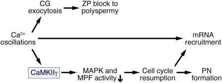
Fertilization triggers a rise in intracellular Ca(2+) concentration ([Ca(2+)](i)) in the egg that initiates a series of events known as egg activation. These events include cortical granule exocytosis that establishes a block to polyspermy, resumption of meiosis, and recruitment of maternal mRNAs into polysomes for translation. Several calcium-dependent proteins, including calcium/calmodulin-dependent protein kinase II (CaMKII), have been implicated in egg activation. However, the precise role of CaMKII in mediating specific events of egg activation and the identity of the isoform(s) present in mouse eggs have not been unequivocally established. Through targeted deletion of the gamma isoform of CaMKII, we find that CaMKIIgamma is the predominant CaMKII isoform in mouse eggs and that it is essential for egg activation. Although CaMKIIgamma(-/-) eggs exhibit a normal pattern of Ca(2+) oscillations after insemination and undergo cortical granule exocytosis, they fail to resume meiosis or to recruit maternal mRNAs. Surprisingly, we find that the recruitment of maternal mRNAs does not directly depend on CaMKII, but requires elevated [Ca(2+)](i) and metaphase II exit. We conclude that CaMKIIgamma specifically controls mouse egg activation by regulating cell cycle resumption.


Conflict of interest statement

The authors declare no conflict of interest.

Figures

Fig. 1.
Fig. 1.
Targeting of the mouse CaMKIIγ gene. (A) CaMKIIγ protein structure, intron–exon structure of the CaMKIIγ gene, and gene targeting strategy. (B) Representative Southern blot of genomic DNA from gene-targeted embryonic stem cells digested with BamHI (BHI) using a probe hybridizing to a genomic region upstream of the long arm of the targeted region. Wild-type (WT) and mutant bands are shown. CaMKIIγ genotypes are shown at the top. (C) RT-PCR to detect CaMKIIγ transcripts in WT and CaMKIIγ−/− (KO) mice. The reverse primer lies in exon 19, and the numbers of the forward primers correspond to the exons containing their sequence.
Fig. 2.
Fig. 2.
CaMKIIγ is the predominant CaMKII isoform in mouse oocytes and eggs. (A) Real-time RT-PCR of CaMKIIα, β, γ, and δ transcripts in mouse oocytes. The experiment was performed three times and a representative example is shown. (B) Lysates from 50 oocytes or eggs obtained from mice of the indicated CaMKIIγ genotypes were resolved on a 10% SDS/PAGE gel that was then subjected to immunoblotting with an antibody directed against the C-termini of all four CaMKII isoforms. CaMKIIγ migrates as a doublet. The experiment was performed three times and a representative immunoblot is shown.
Fig. 3.
Fig. 3.
CaMKIIγ KO female mice are infertile because of a failure to resume meiosis II. (A and B) WT and KO eggs were subjected to SrCl2 activation, and pronuclei (PN) formation was assessed 6–7 h postactivation. Arrowheads indicate the maternal PN observed in WT, but not KO, eggs. (CF) Spindle morphology in WT and KO eggs before (C and D) and after (E and F) SrCl2 activation. Green, β-tubulin; blue, DNA. Representative images are shown. (G) Microinjection of CaMKIIγ or CaMKIIδ cRNA into KO oocytes. As controls, uninjected oocytes (-) and oocytes injected with GFP cRNA were used. Oocytes were matured for 16 h and then SrCl2 activated, and PN formation was determined 6–7 h after activation. The experiment was performed twice. Results are expressed as mean ± range.
Fig. 4.
Fig. 4.
Events of egg activation (EEA) in CaMKIIγ KO eggs. (A) Proteolytic cleavage of ZP2 as a proxy for the postfertilization block to polyspermy. (Upper Left Panel) Protein extracts from WT or KO eggs were prepared before (WT, KO) or 24 h after (fWT, fKO) fertilization and subjected to SDS/PAGE followed by immunoblot with an antibody against ZP2. (Upper Right Panel) Protein extracts from KO eggs were prepared and treated as described for the Left Panel. 0 h, unfertilized metaphase II eggs; fKO, KO female mice were mated to males and eggs were collected at ∼24 h after fertilization (36 h after hCG); 24h, eggs collected at the same time point from unmated females. (Lower Panel) WT and KO eggs were parthenogenetically activated using 10 mM SrCl2. −, eggs cultured without SrCl2; +, eggs cultured with SrCl2. The experiment was performed three times and a representative example is shown. (B) MPF and MAPK assays of metaphase II eggs before (WT, KO) and 12 h after (fWT, fKO) fertilization. Kinase activities were measured in single eggs and are expressed relative to unfertilized WT eggs. Solid bars, MPF activity; open bars, MAPK activity. Data are expressed as the mean ± SEM of five experiments. *P < 0.001, one-way ANOVA. (C) Changes in protein synthesis in metaphase II eggs after fertilization as assessed by [35S]-methionine metabolic radiolabeling and SDS/PAGE. Eggs were isolated from WT and KO mice before (0 h) or 9 h after (fert) fertilization. As a control, eggs were isolated at the same time from unmated females (9h). The experiment was conducted three times and a representative autoradiogram is shown. (D) Changes in protein synthesis in metaphase II eggs after parthenogenetic activation. Metaphase II eggs were isolated from WT and KO mice and treated with SrCl2 (Sr2+), roscovitine (Rosc), or SrCl2 + roscovitine. The experiment was conducted twice and a representative autoradiogram is shown. Bracket depicts 35-kDa complex; arrow indicates position of spindlin (C and D).
Fig. 5.
Fig. 5.
Working model of mouse egg activation. A rise in [Ca2+]i activates the γ isoform of CaMKII that in turn triggers meiotic resumption by decreasing MAPK and MPF activity. Cortical granule (CG) exocytosis and the subsequent ZP block to polyspermy are triggered by [Ca2+]i in a CaMKII-independent manner. Maternal mRNA recruitment is elicited by [Ca2+]i and requires cell cycle resumption but does not directly depend on CaMKII.

References

    1. Schultz RM, Kopf GS. Molecular basis of mammalian egg activation. Curr Top Dev Biol. 1995;30:21–62. - PubMed
    1. Stricker SA. Comparative biology of calcium signaling during fertilization and egg activation in animals. Dev Biol. 1999;211:157–176. - PubMed
    1. Swann K, Saunders CM, Rogers NT, Lai FA. PLCzeta(zeta): A sperm protein that triggers Ca2+ oscillations and egg activation in mammals. Semin Cell Dev Biol. 2006;17:264–273. - PubMed
    1. Cuthbertson KS, Whittingham DG, Cobbold PH. Free Ca2+ increases in exponential phases during mouse oocyte activation. Nature. 1981;294:754–757. - PubMed
    1. Miyazaki S, Igusa Y. Fertilization potential in golden hamster eggs consists of recurring hyperpolarizations. Nature. 1981;290:702–704. - PubMed

Publication types

LinkOut - more resources