

doi: 10.1038/nsmb.1705.
Epub 2009 Dec 6.
Helicobacter pylori CagA inhibits PAR1-MARK family kinases by mimicking host substrates
Affiliations
- PMID: 19966800
- PMCID: PMC3006182
- DOI: 10.1038/nsmb.1705
Item in Clipboard
Helicobacter pylori CagA inhibits PAR1-MARK family kinases by mimicking host substrates
Nat Struct Mol Biol.
2010 Jan.
Abstract
The CagA protein of Helicobacter pylori interacts with numerous cellular factors and is associated with increased virulence and risk of gastric carcinoma. We present here the cocrystal structure of a subdomain of CagA with the human kinase PAR1b/MARK2, revealing that a CagA peptide mimics substrates of this kinase family, resembling eukaryotic protein kinase inhibitors. Mutagenesis of conserved residues central to this interaction renders CagA inactive as an inhibitor of MARK2.
Conflict of interest statement
The authors declare that they have no competing financial interests.
Figures
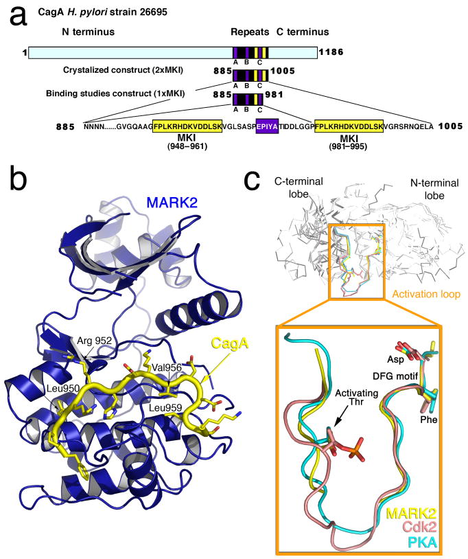
Overall Structure of the CagA-MARK2 Complex. (a) Schematic representation of CagA. The A, B, and C EPIYA sequence repeats are shown as blue boxes. The crystallized construct (885-1005) and the deletion mutant used in binding studies that lacks one of the MKI sequences (885-981) are shown schematically as well. (b) Ribbon diagram of CagA-MARK2 complex, with MARK2 in blue, and the ordered MARK2 inhibitory sequence (MKI, MARK2 Kinase Inhibitor, residues 948-961 and 982-995), shown in yellow. (c) Alignement of several protein kinases, focusing on the activation loop. Cdk2 (PDB ID 1JST) and PKA (PDB ID 1ATP) are from structures of the kinases in activated states (including Cdk2 bound to cyclinA with activating phosphorylation of threonine).
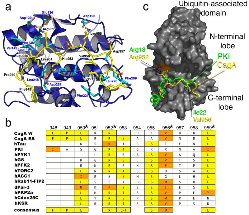
CagA is a Pathogenic Mimic of Host Substrates. (a) Details of the CagA peptide interaction. MARK2 in blue with cyan side-chains, while the MKI peptide of CagA in yellow. (b) Alignment of PAR1/MARK and AMPK family substrates with CagA peptide and, for comparison, PKI. Consensus identity is highlighted in yellow, and conservation in orange. Φ indicates a hydrophobic consensus. CagAW is CagA from H. pylori 26695 (Western subtype); CagA EA is Eastern-Asian subtype of CagA. (c) Superposition of PKI/CagA obtained from aligning the kinases PKA and MARK2. The surface of MARK2 is shown in dark grey. Glu136 of MARK2, which forms a salt bridge with CagA Arg952, is shown in orange on the surface of MARK2.
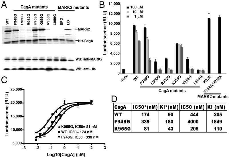
Mutational analysis of MKI mutants. (a) Binding of wild type or mutant hexahistidine-tagged CagA(885-981) to wild type or mutant MARK2(39-364) was assayed by pull-down experiments on Ni-NTA sepharose columns. Eluted material was subjected to SDS-PAGE and proteins stained with coomassie blue dye. Expression levels of MARK2 were determined by Western blot of total cell extract with anti-MARK2 antibody, and anti-His antibody for CagA expression. WT-wild type; EFD-MARK2 mutant (E136G, F138G, D139G); LD-MARK2 mutant (L248G, D251G). (b) Kinase activity of MARK2 in the presence of wild type or mutant CagA synthetic MKI peptides using a luminescent kinase assay. TR1 tau peptide (NVKSKIGSTENLK) at 500μM was used as a substrate with three different concentrations (100μM, 10μM, and 1μM) of CagA peptide inhibitors. MARK2 kinase-inactivated mutants, K82R and T208A/S212A, are negative controls. Error bars represent standard deviations from the mean. (c) Determination of IC50 and Ki of CagA peptide inhibitors of MARK2 through a luminescence-based kinase assay. *-labeled values (IC50* and Ki*) refer to kinase assays in which MARK2 was activated with MARKK. TR1 tau peptide was used as substrate (150μM- in the experiments with previously activated MARK2 or 200μM- in experiments without prior activation of MARK2), with increasing concentration of inhibitory synthetic CagA peptides. Error bars represent standard deviations from the mean. (d) Table summarizing IC50 and Ki data.
References
Publication types
MeSH terms
Substances
Associated data
- Actions
Grants and funding
LinkOut - more resources
Full Text Sources
Other Literature Sources
Molecular Biology Databases
