. 2010 Jan;16(1):59-66.
doi: 10.1038/nm.2063. Epub 2009 Nov 22.

A p53-dependent mechanism underlies macrocytic anemia in a mouse model of human 5q- syndrome

Affiliations

A p53-dependent mechanism underlies macrocytic anemia in a mouse model of human 5q- syndrome

Jillian L Barlow et al. Nat Med. 2010 Jan.

Abstract

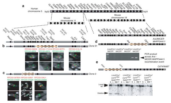
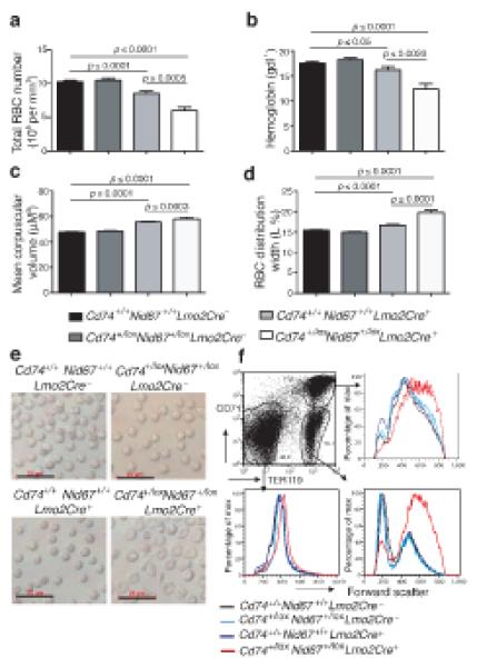
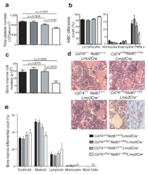
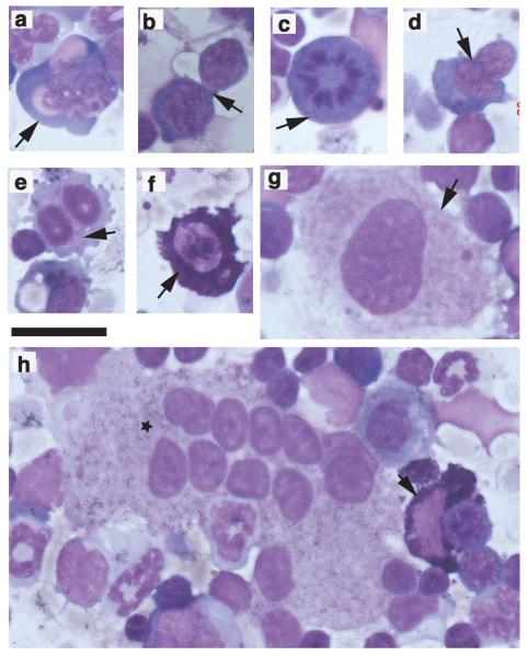
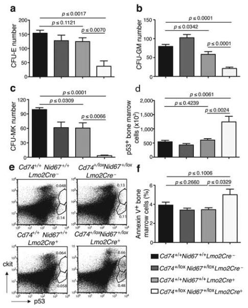
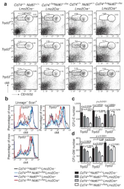
The identification of the genes associated with chromosomal translocation breakpoints has fundamentally changed understanding of the molecular basis of hematological malignancies. By contrast, the study of chromosomal deletions has been hampered by the large number of genes deleted and the complexity of their analysis. We report the generation of a mouse model for human 5q- syndrome using large-scale chromosomal engineering. Haploinsufficiency of the Cd74-Nid67 interval (containing Rps14, encoding the ribosomal protein S14) caused macrocytic anemia, prominent erythroid dysplasia and monolobulated megakaryocytes in the bone marrow. These effects were associated with defective bone marrow progenitor development, the appearance of bone marrow cells expressing high amounts of the tumor suppressor p53 and increased bone marrow cell apoptosis. Notably, intercrossing with p53-deficient mice completely rescued the progenitor cell defect, restoring common myeloid progenitor and megakaryocytic-erythroid progenitor, granulocyte-monocyte progenitor and hematopoietic stem cell bone marrow populations. This mouse model suggests that a p53-dependent mechanism underlies the pathophysiology of the 5q- syndrome.


Figures

Figure 1
Figure 1
Representation of human 5q syndrome CDR in humans and alignment with mouse regions of synteny. (a) The human 5q syndrome CDR aligned with mouse chromosomes 11 and 18. (b) Confirmation of Clone 91 using FISH. (c) Confirmation of Clone 2 using FISH. (d) Confirmation of the Arsi – micerMHPP344i11 deletion by PCR analysis (see also Supplementary Fig. 3). (e) Confirmation of the Cd74Nid67 deletion by Southern analysis (see also Supplementary Fig. 4). S, spleen; L, liver; BM, bone marrow; T, tail. WT, wildtype. Arrows indicate the transcriptional orientation of the genes. The BAC clones and dye colours (red or green) used for FISH are marked in their relative positions.
Figure 2
Figure 2
Deletion of the Cd74+/loxNid67+/lox interval leads to macrocytic anemia. Blood from the specific genotypes was analysed (n = 28 – 52). (a) Red blood cell numbers. (b) Hemoglobin levels. (c) Mean corpuscular volume (MCV). (d) Red cell distribution width (RDW) (n = 28 – 52). (e) Live RBC imaged using light microscopy. Scale bar, 20 μm. (f) Bone marrow cells gated on CD71 and TER119 expression and analysed for forward scatter as a measure of cell size. Data are representative of two experiments with five mice per group.
Figure 3
Figure 3
Blood and bone marrow cell proportions vary following deletion of the Cd74+/loxNid67+/lox interval. (a) Platelet numbers in blood (n = 28 – 52). (b) Differential white blood cell counts (n = 5 per group). (c) Bone marrow cell numbers (n = 5 per group). Data are representative of at least two experiments. (d) Hematoxylin and eosin stained bone marrow sections. Scale bar, 20 μm. Representative of five mice per group all of similar age. (e) Bone marrow differential cell counts from cytospins (n = 8 - 10 per group).
Figure 4
Figure 4
Giemsa-stained bone marrow cytospins following deletion of the Cd74+/loxNid67+/lox interval to show prominent dyserythropoiesis, megakaryocytes with monolobulated nuclei and mast cells. Scale bar 40 μm. (a) dysplastic erythroblast (arrow); (b) inter-cytoplasmic bridging (arrow); (c) mitotic figure (arrow); (d) inter-nuclear bridging (arrow); (e) binucleate erythroid precursors (arrow); (f) mast cell (arrow); (g) megakaryocyte with monolobulated nucleus (arrow); (h) megakaryocyte with multiple monolobulated nuclei (star). Also includes a mast cell (arrow).
Figure 5
Figure 5
Hematopoietic cell progenitors and p53 positive and apoptotic cells in the Cd74+/loxNid67+/lox, Lmo2Cre+ mice. (a) Number of colony forming units – erythroid in bone marrow cultures (n = 5 per group). Data are representative of two experiments. (b) Number of colony forming units – granulocyte/macrophage in bone marrow cultures (n = 7 per group). (c) Number of colony forming units – megakaryocyte in bone marrow cultures (n = 3 per group). (d) Numbers of p53+ bone marrow cells (n = 5 per group). Representative of two similar experiments. (e) Intracellular staining for p53 in cKit+ cell surface stained cells. Example is of a highly p53-positive Cd74+/loxNid67+/lox, Lmo2Cre+, sample to show cKit+ cell involvement. (f) Annexin V staining of bone marrow cells (n = 5 per group).
Figure 6
Figure 6
Deletion of p53 reverses the progenitor cell deficits that result from the deletion of the Cd74+/loxNid67+/lox interval. (a) Bone marrow cells stained for CMP, MEP and GMP cell surface markers (representative of five mice per group). (b) Bone marrow cells stained for HSC cell surface markers. Cells initially gated on LinSca1+ (representative of five mice per group). (c) Number of colony forming units – erythroid in bone marrow cultures (n = 6 per group). (d) Number of colony forming units – granulocyte/macrophage in bone marrow cultures (n = 6 per group).

Comment in

References

    1. Van den Berghe H, et al. Distinct hematological disorder with deletion of long arm of no. 5 chromosome. Nature. 1974;251:437–438. - PubMed
    1. Giagounidis AA, Germing U, Wainscoat JS, Boultwood J, Aul C. The 5q- syndrome. Hematology. 2004;9:271–277. - PubMed
    1. List A, et al. Lenalidomide in the myelodysplastic syndrome with chromosome 5q deletion. N Engl J Med. 2006;355:1456–1465. - PubMed
    1. Vardiman JW, Harris NL, Brunning RD. The World Health Organization (WHO) classification of the myeloid neoplasms. Blood. 2002;100:2292–2302. - PubMed
    1. Boultwood J, Lewis S, Wainscoat JS. The 5q-syndrome. Blood. 1994;84:3253–3260. - PubMed

Publication types