

A mitotic transcriptional switch in polycystic kidney disease
- PMID: 19966811
- PMCID: PMC3062536
- DOI: 10.1038/nm.2068
A mitotic transcriptional switch in polycystic kidney disease
Abstract
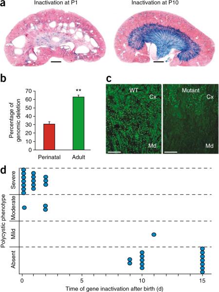
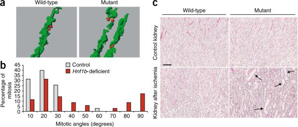
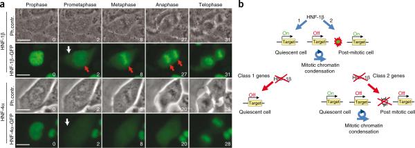
Hepatocyte nuclear factor-1beta (HNF-1beta) is a transcription factor required for the expression of several renal cystic genes and whose prenatal deletion leads to polycystic kidney disease (PKD). We show here that inactivation of Hnf1b from postnatal day 10 onward does not elicit cystic dilations in tubules after their proliferative morphogenetic elongation is over. Cystogenic resistance is intrinsically linked to the quiescent state of cells. In fact, when Hnf1b deficient quiescent cells are forced to proliferate by an ischemia-reperfusion injury, they give rise to cysts, owing to loss of oriented cell division. Remarkably, in quiescent cells, the transcription of crucial cystogenic target genes is maintained even in the absence of HNF-1beta. However, their expression is lost as soon as cells proliferate and the chromatin of target genes acquires heterochromatin marks. These results unveil a previously undescribed aspect of gene regulation. It is well established that transcription is shut off during the mitotic condensation of chromatin. We propose that transcription factors such as HNF-1beta might be involved in reprogramming gene expression after transcriptional silencing is induced by mitotic chromatin condensation. Notably, HNF-1beta remains associated with the mitotically condensed chromosomal barrels. This association suggests that HNF-1beta is a bookmarking factor that is necessary for reopening the chromatin of target genes after mitotic silencing.
Figures

Similar articles
-
Role of transcription factor hepatocyte nuclear factor-1β in polycystic kidney disease.Cell Signal. 2020 Jul;71:109568. doi: 10.1016/j.cellsig.2020.109568. Epub 2020 Feb 14. Cell Signal. 2020. PMID: 32068086 Free PMC article. Review.
-
HNF-1beta regulates transcription of the PKD modifier gene Kif12.J Am Soc Nephrol. 2009 Jan;20(1):41-7. doi: 10.1681/ASN.2008020238. Epub 2008 Nov 12. J Am Soc Nephrol. 2009. PMID: 19005009 Free PMC article.
-
Hepatocyte nuclear factor 1β suppresses canonical Wnt signaling through transcriptional repression of lymphoid enhancer-binding factor 1.J Biol Chem. 2020 Dec 18;295(51):17560-17572. doi: 10.1074/jbc.RA120.015592. J Biol Chem. 2020. PMID: 33453998 Free PMC article.
-
New insights into the role of HNF-1β in kidney (patho)physiology.Pediatr Nephrol. 2019 Aug;34(8):1325-1335. doi: 10.1007/s00467-018-3990-7. Epub 2018 Jul 1. Pediatr Nephrol. 2019. PMID: 29961928 Free PMC article. Review.
-
Mechanism of Fibrosis in HNF1B-Related Autosomal Dominant Tubulointerstitial Kidney Disease.J Am Soc Nephrol. 2018 Oct;29(10):2493-2509. doi: 10.1681/ASN.2018040437. Epub 2018 Aug 10. J Am Soc Nephrol. 2018. PMID: 30097458 Free PMC article.
Cited by
-
HNF1B Transcription Factor: Key Regulator in Renal Physiology and Pathogenesis.Int J Mol Sci. 2024 Oct 2;25(19):10609. doi: 10.3390/ijms251910609. Int J Mol Sci. 2024. PMID: 39408938 Free PMC article. Review.
-
Hedgehog signaling is required for the maintenance of mesenchymal nephron progenitors.bioRxiv [Preprint]. 2024 May 5:2023.08.12.553098. doi: 10.1101/2023.08.12.553098. bioRxiv. 2024. PMID: 37645929 Free PMC article. Preprint.
-
GATA2 mitotic bookmarking is required for definitive haematopoiesis.Nat Commun. 2023 Aug 14;14(1):4645. doi: 10.1038/s41467-023-40391-x. Nat Commun. 2023. PMID: 37580379 Free PMC article.
-
Generation of c-Myc transgenic pigs for autosomal dominant polycystic kidney disease.Transgenic Res. 2013 Dec;22(6):1231-9. doi: 10.1007/s11248-013-9707-6. Epub 2013 Mar 30. Transgenic Res. 2013. PMID: 23543409
-
Functional genomics analysis identifies loss of HNF1B function as a cause of Mayer-Rokitansky-Küster-Hauser syndrome.Hum Mol Genet. 2023 Mar 6;32(6):1032-1047. doi: 10.1093/hmg/ddac262. Hum Mol Genet. 2023. PMID: 36282544 Free PMC article.
References
-
- Gottesfeld JM, Forbes DJ. Mitotic repression of the transcriptional machinery. Trends Biochem. Sci. 1997;22:197–202. - PubMed
-
- Torres VE, Harris PC, Pirson Y. Autosomal dominant polycystic kidney disease. Lancet. 2007;369:1287–1301. - PubMed
-
- Fischer E, Gresh L, Reimann A, Pontoglio M. Cystic kidney diseases: learning from animal models. Nephrol. Dial. Transplant. 2004;19:2700–2702. - PubMed
Publication types
MeSH terms
Substances
Grants and funding
LinkOut - more resources
Full Text Sources
Other Literature Sources
Molecular Biology Databases
