. 2010 Mar 11;29(10):1486-97.
doi: 10.1038/onc.2009.443. Epub 2009 Dec 7.

Depletion of WRN protein causes RACK1 to activate several protein kinase C isoforms

Affiliations

Depletion of WRN protein causes RACK1 to activate several protein kinase C isoforms

L Massip et al. Oncogene. .

Abstract

Werner's syndrome (WS) is a rare autosomal disease characterized by the premature onset of several age-associated pathologies. The protein defective in patients with WS (WRN) is a helicase/exonuclease involved in DNA repair, replication, transcription and telomere maintenance. In this study, we show that a knock down of the WRN protein in normal human fibroblasts induces phosphorylation and activation of several protein kinase C (PKC) enzymes. Using a tandem affinity purification strategy, we found that WRN physically and functionally interacts with receptor for activated C-kinase 1 (RACK1), a highly conserved anchoring protein involved in various biological processes, such as cell growth and proliferation. RACK1 binds strongly to the RQC domain of WRN and weakly to its acidic repeat region. Purified RACK1 has no impact on the helicase activity of WRN, but selectively inhibits WRN exonuclease activity in vitro. Interestingly, knocking down RACK1 increased the cellular frequency of DNA breaks. Depletion of the WRN protein in return caused a fraction of nuclear RACK1 to translocate out of the nucleus to bind and activate PKCdelta and PKCbetaII in the membrane fraction of cells. In contrast, different DNA-damaging treatments known to activate PKCs did not induce RACK1/PKCs association in cells. Overall, our results indicate that a depletion of the WRN protein in normal fibroblasts causes the activation of several PKCs through translocation and association of RACK1 with such kinases.


Conflict of interest statement

Conflict of interest

The authors declare no conflict of interest.

Figures

Figure 1
Figure 1
Impact of WRN protein depletion on GM08402 diploid fibroblasts cell cycle and phospho-proteins after transfection with scrambled siRNA and WRN-specific siRNA. (a) WRN protein detection with an anti-WRN antibody (Santa Cruz Biotechnology, Santa Cruz, CA, USA) in GM08402 cells 48 h after siRNA molecule transfection. The β-actin protein present in the lysate was used as control. (b) Percentage of GM08402 cells (at passage 8) in each phase of the cell cycle 48 h after transfection with control siRNA and siWRN molecules determined by FACS analysis. Experiments were performed in duplicate. (c) Detergent-solubilized lysates from control siRNA- and siWRN-transfected fibroblasts were subjected to Kinetworks custom multi-sample screen (KCSS-1.0) analyses (http://www.Kinexus.ca). Antibodies used in this study (from Kinexus Bioinformatics Corp.) were against phosphothreonine 232 of FOS, phospho-serines 21 of GSK3α/β, IKKβ, phosphoserine 729 of PKCε, PKCλ/ι and phospho-serine 910 of PKD. WRN, protein defective in patients with Werner’s syndrome; siRNA, small interfering RNA; FACS, fluorescence-activated cell sorting; PKC, protein kinase C.
Figure 2
Figure 2
Interaction of RACK1 with the WRN protein. (a) Coprecipitation of RACK1 with the TAP-WRN protein in HT1080 fibrosarcoma cells. Proteins from TAP and TAP-WRN expressing cells were eluted from the streptavidin beads and analyzed by SDS–PAGE with antibodies against WRN and RACK1 proteins. (b) Coimmunoprecipitation of human WRN protein with RACK1. Approximately 2 mg of proteins from human HT1080 cells were immunoprecipitated with antibodies against the N- or C-terminus region of the human WRN protein. Control antibodies were of the same IgG species. Immunoprecipitates were analyzed by western blotting with the anti-WRN antibody (WRN; top panel) and an antibody against RACK1. Proteins were revealed with an ECL kit. The anti-WRN (N-ter) antibody is from Novus Biologicals (Littleton, CO, USA). The anti-WRN (C-ter) and anti-RACK1 antibodies are from Santa Cruz Biotechnology. The ‘input’ lane corresponds to 20 μg of total cell lysate. (c) Binding of purified WRN protein to GST-RACK1 affinity Sepharose beads as revealed by immunoblots with an anti-WRN antibody. GST Sepharose beads were used as a negative control. The experiment is presented in duplicate. (d) Interaction of RACK1 with different domains of WRN in whole-cell extract. Immunoblot against RACK1 protein bound to different GST-WRN affinity Sepahrose beads. Human HT1080 whole-cell extracts were incubated with 50 μg of the GST-WRN fragments or GST-linked glutathione-Sepharose beads overnight. Proteins bound to the affinity beads were analyzed by SDS–PAGE with antibodies against RACK1. (e) Schematic representation of different WRN fragments that were used in the WRN affinity chromatography experiments. Each domain of the WRN protein is indicated on the full WRN protein figure. The amino-acid residues of the WRN fragments used in this study are indicated on the top of each construct. Binding of RACK1 is indicated on the right by the ‘+’ sign. The ‘−’ sign indicates no binding detected. RACK1, receptor for activated C-kinase 1; TAP, tandem affinity purification; WRN, protein defective in patients with Werner’s syndrome.
Figure 3
Figure 3
The impact of purified RACK1 on DNA helicase and exonuclease activities of purified WRN protein, and the impact of RACK1 depletion on DNA damage in cells. (a) The indicated concentration of purified human RACK1 protein was incubated with 3nM of purified WRN protein, and the indicated radioactive DNA substrate under standard conditions for helicase activity for 30 min at 37 °C. Reactions were stopped in the appropriate dye buffer, and DNA products were run on a 12% native polyacrylamide gel. The double- and single-stranded DNA structures are depicted on the left of the autoradiogram. The 5′-labeled strand of the duplex is represented by an asterisk (*). The triangle represents heat-denatured DNA. (b) The indicated concentrations of purified human RACK1 and WRN proteins were incubated with a radioactive DNA-forked structure under the same buffer conditions as for the helicase assay for 30 min at 37 °C. Reactions were stopped in the appropriate dye buffer, heat denatured, and the DNA substrates were analyzed on a 12% denaturing gel. (c) The percentage of cells displaying foci of DNA damage detected by the anti-γ-H2AX antibody. The percentage of cells exhibiting 0–10 foci, 11–20 foci or >20 foci is depicted for each transfection experiment (150 cells per transfection were analyzed). The resulting contingency table is displayed as a histogram. (d) Extent of DNA breaks quantified by the alkaline comet assay. The tails of broken DNA from 100 cells were measured to obtain the mean tail length (in μm). All experiments were repeated twice (unpaired Student’s t-test: *P=0.049; **P<0.001. Bars represent s.e.m.). RACK1, receptor for activated C-kinase 1; WRN, protein defective in patients with Werner’s syndrome.
Figure 4
Figure 4
Nuclear RACK1 colocalizes with nucleoplasmic WRN protein in normal and tumor cells. Immuno-localization of RACK1 and WRN proteins in HT1080 fibrosarcoma cells is shown in the top row. The bottom row shows immuno-localization of RACK1 and WRN proteins in normal GM08402 fibroblasts. Anti-mouse Alexa-594-labeled and anti-rabbit Alexa-488-conjugated secondary antibodies were used to visualize WRN and RACK1 by confocal microscopy at 568 and 488 nm, respectively. Images depict representative cells at ×600. In the merged images, a yellow color appears where RACK1 (red) and WRN (green) fluorescence signals coincide. RACK1, receptor for activated C-kinase 1; WRN, protein defective in patients with Werner’s syndrome.
Figure 5
Figure 5
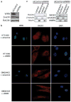
The impact of WRN depletion on RACK1 in HT1080 and GM08402 cells. (a) Expression levels of total WRN and RACK1 proteins in control siRNA- and siWRN-transfected HT1080 cells. The β-actin protein is used as loading control. (b) RACK1 protein localization detected by immunofluorescence in HT1080 and GM08402 cells transfected with control siRNA or siWRN molecules. (c) Expression levels of RACK1 proteins in the membrane fraction of control siRNA- and siWRN-transfected HT1080 cells (panels on the left). TRAP1 protein was used as loading control for the membrane fraction (including mitochondrial membranes). β-Tubulin was used as a control for cytosolic fractions. Expression levels of RACK1 proteins in the nuclear and cytoplasmic fractions of control siRNA- and siWRN-transfected HT1080 cells (panels on the right). β-Tubulin was used as a control for the cytoplasmic fraction. Heterogeneous nucleoriboprotein K (HNRPK) was used as a control for the nuclear fraction. RACK1, receptor for activated C-kinase 1; WRN, protein defective in patients with Werner’s syndrome; siRNA, small interfering RNA.
Figure 6
Figure 6
The impact of WRN protein depletion on RACK1/PKC associations. (a) Coprecipitation of three different protein kinase C with the TAP-RACK1 protein in HT1080 fibrosarcoma cells. Proteins from TAP and TAP-RACK1 expressing cells were eluted from the streptavidin beads and analyzed by SDS–PAGE with antibodies against PKCδ, PKCε and PKCβII. (b) Coprecipitation of two phosphorylated PKC with the TAP-RACK1 protein in HT1080 fibrosarcoma cells. Proteins from TAP and TAP-RACK1 expressing cells were eluted from the streptavidin beads and analyzed by SDS–PAGE with antibodies against phospho-threonine 507 of PKCδ and phospho-threonine 641 of PKCβII. TAP-RACK1 is shown in the top panel with an anti-RACK1 antibody. RACK1, receptor for activated C-kinase 1; PKC, protein kinase C; WRN, protein defective in patients with Werner’s syndrome; TAP, tandem affinity purification.
Figure 7
Figure 7
The impact of RACK1 on the phosphorylation of PKCδ, PKCβII and PKCε in WRN-depleted GM08402 cells. Immunoblot analyses with antibodies against WRN, RACK1, PKCδ, phospho-threonine 507 of PKCδ, total PKCδ, phosphothreonine 641 of PKCβII, total PKCβII, phospho-serine 729 of PKCε, and total PKCε 48 h after GM08402 cells were transfected with control-scrambled siRNA, and siWRN with or without siRACK1 molecules. RACK1, receptor for activated C-kinase 1; PKC, protein kinase C; WRN, protein defective in patients with Werner’s syndrome; siRNA, small interfering RNA.
Figure 8
Figure 8
The impact of RACK1 protein knock down on reactive oxygen species (ROS) production and senescence markers in WRN-depleted normal human GM08402 fibroblasts. (a) RACK1 protein levels in GM08402 fibroblasts 48 h after transfection with control-scrambled siRNA or siRACK1 molecules. β-actin is used as control. (b) PKCδ protein levels in GM08402 fibroblasts 48 h after transfection with control-scrambled siRNA or siPKCδ molecules. RACK1 protein level is used as a control. (c) Intracellular ROS levels in GM08402 cells transfected with control siRNA (siControl), siWRN, siWRN+siRACK1 or siWRN+siPKCδ molecules in combination. Experiments were performed in triplicates (unpaired Student’s t-test; *P<0.05). Bars represent s.e.m.

Similar articles

Cited by

References

    1. Battaini F, Pascale A, Paoletti R, Govoni S. The role of anchoring protein RACK1 in PKC activation in the ageing rat brain. Trends Neurosci. 1997;20:410–415. - PubMed
    1. Beckman KB, Ames BN. The free radical theory of aging matures. Physiol Rev. 1998;78:547–581. - PubMed
    1. Berns H, Humar R, Hengerer B, Kiefer FN, Battegay EJ. RACK1 is up-regulated in angiogenesis and human carcinomas. FASEB J. 2000;14:2549–5258. - PubMed
    1. Besson A, Wilson TL, Yong VW. The anchoring protein RACK1 links protein kinase Cepsilon to integrin beta chains. Requirements for adhesion and motility. J Biol Chem. 2002;277:22073–22084. - PubMed
    1. Brosh RM, Jr, von Kobbe C, Sommers JA, Karmakar P, Opresko PL, Piotrowski J, et al. Werner syndrome protein interacts with human flap endonuclease 1 and stimulates its cleavage activity. EMBO J. 2001;20:5791–5801. - PMC - PubMed

Publication types

MeSH terms