. 2010 Mar 11;29(10):1475-85.
doi: 10.1038/onc.2009.440. Epub 2009 Dec 7.

The IGF-1/IGF-1R signaling axis in the skin: a new role for the dermis in aging-associated skin cancer

Affiliations

The IGF-1/IGF-1R signaling axis in the skin: a new role for the dermis in aging-associated skin cancer

D A Lewis et al. Oncogene. .

Abstract

The appropriate response of human keratinocytes to ultraviolet-B (UVB) is dependent on the activation status of the insulin-like growth factor 1 (IGF-1) receptor. Keratinocytes grown in conditions in which the IGF-1 receptor is inactive inappropriately replicate in the presence of UVB-induced DNA damage. In human skin, epidermal keratinocytes do not express IGF-1, and hence the IGF-1 receptor on keratinocytes is activated by IGF-1 secreted from dermal fibroblasts. We now show that the IGF-1 produced by human fibroblasts is essential for the appropriate UVB response of keratinocytes. Furthermore, the expression of IGF-1 is silenced in senescent fibroblasts in vitro. Using quantitative reverse transcriptase-PCR and immunohistochemisty, we can show that IGF-1 expression is also silenced in geriatric dermis in vivo. The diminished IGF-1 expression in geriatric skin correlates with an inappropriate UVB response in geriatric volunteers. Finally, the appropriate UVB response is restored in geriatric skin in vivo through pretreatment with exogenous IGF-1. These studies provide further evidence for a role of the IGF-1 receptor (IGF-1R) in suppressing UVB-induced carcinogenesis, suggest that fibroblasts have a critical role in maintaining appropriate activation of the keratinocyte IGF-1R, and imply that reduced expression of IGF-1 in geriatric skin could be an important component in the development of aging-related non-melanoma skin cancer.


Figures

Figure 1
Figure 1. Exogenous IGF-1 (but not insulin) regulates the response of human keratinocytes to UVB irradiation
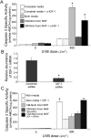
(A) When insulin is withheld from keratinocyte media, keratinocytes become more sensitive to UVB-induced apoptosis. Keratinocytes were grown in EpiLife NoIn media for 16 h. At that time the media was replaced with EpiLife Complete media, EpiLife NoIn media, or EpiLife NoIn media containing the indicated concentrations of IGF-1 or insulin for thirty min and then irradiated with 400 J/m2 of UVB. Keratinocytes were harvested six hours post-irradiation and assayed for the induction of apoptosis by measuring the induction of caspase 3 specific activity. Error bars indicate the standard error of the mean; the asterisks indicate significant difference between caspase 3 specific activities derived from keratinocytes grown in unsupplemented EpiLife NoIn media (p<0.005, t-test). The data presented represent three independent assays. (B) Keratinocytes irradiated in the absence of IGF-1 do not undergo UVB-induced senescence. Keratinocytes were grown in EpiLife Complete or EpiLife NoIn media for 16 h and then irradiated with 100 J/m2 of UVB. Twelve hours post-irradiation, the media on all of the plates was replaced with fresh EpiLife Complete media. Seventy-two hours post-irradiation, the keratinocytes were assayed for the expression of senescence-associated β-galactosidase activity (nuclei of senescent cells stain blue). (C) Keratinocytes lacking IGF-1R activation can replicate UVB-damaged DNA. Keratinocytes were grown in EpiLife NoIn (i, iii, v) or EpiLife Complete (ii, iv, vi) media for 16 h and then irradiated with 100 J/m2 of UVB. At 0 (i, ii), 0.25 (iii, iv) or 24 (v, vi) h post-irradiation, the keratinocytes were fixed and assayed for the expression of Ki67 (appearing green) and the presence of UVB-induced thymidine dimers (appearing red). Cells expressing Ki67 and containing thymidine dimers appear yellow.
Figure 2
Figure 2. IGF-1 secreted by normal human fibroblasts can protect keratinocytes from UVB-induced apoptosis
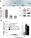
(A) To determine if normal human fibroblasts can produce a protective factor which can activate the IGF-1R and restore the normal UVB response of keratinocytes, normal human fibroblasts were grown in the EpiLife NoIn media for 48 h (referred to as Conditioned Media, CM). Keratinocytes were grown in EpiLife NoIn media for 48 h and subsequently the media was replaced with EpiLife Complete media, EpiLife NoIn media, or EpiLife NoIn-CM derived from fibroblasts. Aliquots of EpiLife Complete media and CM-NoIn were treated with a neutralizing antibody to IGF-1 (Calbiochem, α-IGF-1, Ab2; inhibits 50% at suggested concentration) 15 min prior to adding it to the keratinocytes. One hour following media exchange, the keratinocytes were irradiated with 0 or 400 J/m2 of UVB. Six hours post-irradiation, the keratinocytes were harvested and assayed for the induction of apoptosis. Error bars indicate the standard error of the mean; the asterisk indicates significant difference between caspase 3 specific activities derived from keratinocytes grown in Complete media versus NoIn media (p<0.01, t-test). The data presented represent three independent assays. (B and C) Fibroblasts were treated with IGF-1-specific siRNA or non-specific siRNA for 24 h. NoIn media was then added to the fibroblasts for 48 h. At that time, the media was removed, filter-sterilized, and added to keratinocyte cultures. (B) qRT-PCR analysis of IGF-1 expression in fibroblasts following the indicated siRNA treatment. Error bars indicate the standard error of the mean; asterisk indicates significant (p<0.02, t-test) difference from nonsense siRNA-treated keratinocytes. (C) Keratinocytes were grown in NoIn media for 48 h, then the indicated media was added for 1 h. At that time, the keratinocytes were irradiated with the indicated dose of UVB and harvested six hours post-irradiation for caspase 3 analysis. Error bars indicate the standard error of the mean; the asterisks indicate significant difference between caspase 3 specific activities derived from keratinocytes grown in NoIn media + !GF-1 (p<0.03, t-test). The data presented represent three independent assays.
Figure 3
Figure 3. Induction of senescence (stress-induced or replicative) silences IGF-1 expression in fibroblasts and abolishes the protective effect of conditioned media on UVB-induced apoptosis in keratinocytes
Fibroblasts were treated with 600 μM H2O2 for 2 h and maintained in culture for 72 h (sisNHF) or serially passaged until they reached replicative senescence (rsNHF). (A) Fibroblasts stained for senescence-associated β-galactosidase activity (senescent cells are blue, appearing dark in the image) (B) qRT-PCR analysis of IGF-1 expression. Error bars indicate the standard error of the mean; asterisks indicate significant (p<0.03, t-test) difference from proliferative fibroblasts. (C) Proteins from the indicated cell lysates were immunoblotted and probed with the specified antibodies. (D) Conditioned media from proliferative fibroblasts (NHF-CM) but not from senescent fibroblasts (sisNHF CM or rsNHF-CM) protect keratinocytes from UVB-induced apoptosis. Error bars indicate the standard error of the mean; the asterisks indicate significant difference between caspase 3 specific activities derived from keratinocytes grown in Complete media (p<0.005, t-test). The data presented represent three independent assays.
Figure 4
Figure 4. Decreased expression of IGF-1 in dermal fibroblasts and a corresponding decrease in IGF-1R activation in epidermal keratinocytes in geriatric skin
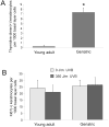
(A) Skin from sun-protected areas of young adult (20-28 years old, panels i, ii) or geriatric (≥65 years old, panels iii, iv) were stained with antibodies specific to human IGF-1 and visualized by anti-rabbit-HRP/DAB staining (positive staining indicated by brown precipitate). The slides were counterstained with hematoxylin. IGF-1 expression is readily observed in individual dermal fibroblasts from young skin (black arrows) but not in geriatric skin (white arrows). (B) Total RNA was extracted from biopsies of skin derived from young adult and geriatric skin. The relative amount of IGF-1 mRNA was determined by qRT-PCR analysis standardized to actin mRNA levels. Error bars indicate the standard error of the mean; asterisk indicates significant (p<0.01, t-test) difference between the IGF-1 mRNA levels from young adult skin versus geriatric skin. (C) Skin from sun-protected areas of young adult (20-28 years old, top panels) or geriatric (≥65 years old, bottom panels) were stained with antibodies specific to activated (tyrosine-phosphorylated) IGF-1R and visualized by anti-rabbit-HRP/DAB staining (positive staining indicated by brown precipitate). The slides were counterstained with hematoxylin. High power micrographs indicate activated IGF-1R expression in nuclei and in discrete pockets at the cell surface (examples indicated by white arrows) of individual epidermal keratinocytes in young skin but not in geriatric skin.
Figure 5
Figure 5. Deficient IGF-1 expression in geriatric fibroblasts increases mutagenic potential in keratinocytes following UVB irradiation
(A) Example of UVB-response assay. A 2 cm2 area on the lower back of a 75 year old volunteer was irradiated with UVB (350 J/m2). Twenty-four hours following irradiation, a four mm punch biopsy was obtained from the irradiated skin. Sections of formalin-fixed, paraffin-embedded tissue were stained with both α-Ki67 and α-thymidine dimer antibodies (panels i, iv: α-Ki67 immunofluorescence only (staining green); panels ii, v: α-thymidine dimer immunofluorescence only (staining red); panels iii, vi: α-Ki67 and α-thymidine dimer double-immunofluorescence (double-positive cells staining yellow). Arrows in i, ii, iii and iv, v, vi indicate the location of the same cell(s) in each of the respective panels. The top series of panels is an example of a cell positive for both Ki67 and thymidine dimmers (staining yellow, inappropriate UVB response) while the cell indicated in bottom panels is only positive for Ki67 (staining green, appropriate UVB response). The location of the basement membrane is indicated by a grey dashed line. (B) Geriatric keratinocytes respond inappropriately to UVB irradiation. A 2 cm2 area on the lower back of young adult (20-28 years old; YA) or geriatric (> 65 years old; GA) volunteers were irradiated with UVB (350 J/m2). Twenty-four hours following irradiation, a four mm punch biopsy was obtained from the irradiated skin and a three mm punch biopsy was removed from unirradiated skin. The tissues were processed as described in (A). The number of basal layer keratinocytes positive for only Ki67, positive for only TD, or positive for both Ki67 and TD were determined for the entire length of each biopsy. Similar sections were stained with H&E to determine the total number of basal layer keratinocytes in each biopsy. The graph represents the number of replicating keratinocytes in the UVB-irradiated skin that contains UVB-induced DNA damage per 1000 basal layer cells. The results presented were derived from six young adult and six geriatric volunteers. Asterisk indicates a statistically significant difference between the two cohorts (p<0.0001, student t-test).
Figure 6
Figure 6. Young adult keratinocytes repair UVB-induced DNA damage more quickly than geriatric keratinocytes
(A) Experiment was conducted as described in Figure 5. The graph represents the total number of keratinocytes in the UVB-irradiated skin that contains UVB-induced DNA damage per 1000 basal layer cells from each treatment group. The results presented were derived from six young adult and seven geriatric volunteers. Asterisks indicates a statistically significant difference between the young adult and geriatric cohorts (p<0.0001, student t-test). (B) No difference between the number of replicating keratinocytes in young adult or geriatric skin. Experiment was conducted as described in Figure 5. The graph represents the total number of replicating keratinocytes per 1000 basal layer cells from each treatment group. There was no statistically significant difference between and of the treatment groups.
Figure 7
Figure 7. Exogenous IGF-1 restores appropriate UVB-response to geriatric skin
(A) Intradermal injection of IGF-1 activates the epidermal IGF-1R. In vitro explants of full-thickness human skin were analyzed for the activation of the IGF-1R using an antibody specific for tyrosine-phosphorylated IGF-1R. i.) Control untreated explanted skin; ii.) IGF-1 (200 ng in 50 μl of saline) was injected into the dermis of explanted human skin 30 minutes prior to harvesting the skin; iii.) AG 538 (a specific small molecule inhibitor or the IGF-1R) was topically applied to the explanted human skin for 30 min prior to sub-dermal injection of IGF-1 (as described in ii). White arrows indicate brown punctate cell membrane staining and black arrows indicate positively-staining brown nuclei. (B) Exogenous IGF-1 corrects the inappropriate UVB-response in geriatric skin. Isolated areas on the lower backs of geriatric volunteers were injected with 50 μl of saline and 50 μl of saline containing 200 ng of IGF-1 just beneath the epidermis. Thirty minutes following the injections, each site was irradiated with 350 J/m2 of UVB. At twenty-four hours post-irradiation, biopsies were obtained from each irradiated site and from an unirradiated site. The tissues were processed as described in (Figure 5b). The results presented in the graph were derived from seven geriatric volunteers. Asterisk indicates a statistically significant difference between the saline and IGF-1 treated sites (p<0.0001; student t-test).
Figure 8
Figure 8. The influence of aging on IGF-1 expression in the skin and its role in UVB-induced carcinogenesis
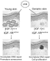
Keratinocytes in aged epidermis exposed to UVB wavelengths in sunlight may respond inappropriately to the UVB exposure. The dermis of young adults produces sufficient levels of IGF-1 to activate the IGF-1R on epidermal keratinocytes. The appropriate activation of the IGF-1R on keratinocytes leads to the induction of stress-induced senescence following sufficient UVB exposure. In contrast, the expression of IGF-1 is silenced in aged dermis. The consequence of diminished IGF-1 expression is a lack of IGF-1R activation in epidermal keratinocytes. Instead of undergoing stress-induced senescence, the aged keratinocytes are able to proliferate in the presence of UVB-damaged DNA. We hypothesize this decrease in IGF-1 expression with advancing age is a contributor to the increase in non-melanoma skin cancer seen in geriatric patients.

References

    1. Chuang T-Y, Lewis DA, Spandau DF. Decreased incidence of nonmelanoma skin cancer in patients with type 2 diabetes mellitus using insulin: a pilot study. Br J Dermatol. 2005;153:552–557. - PubMed
    1. Cotton J, Spandau DF. Ultraviolet B dose influences the induction of apoptosis and p53 in human keratinocytes. Radiat Res. 1997;147:148–155. - PubMed
    1. Dimri GP, Lee X, Basile G, Acosta M, Scott G, et al. A biomarker that identifies senescent human cells in culture and in aging skin in vivo. Proc Natl Acad Sci USA. 1995;92:9363–9367. - PMC - PubMed
    1. Donovan EA, Kummar S. Role of the insulin-like growth factor-1R system in colorectal carcinogenesis. Critical Rev Oncol-Hematol. 2008;66:91–98. - PMC - PubMed
    1. Dziadziuszko R, Camidge DR, Hirsch FR. The insulin-like growth factor in lung cancer. J Thoracic Oncol. 2008;3:815–818. - PubMed

Publication types

MeSH terms

Substances