

Characterization of ionic currents in human neural stem cells
- PMID: 19967046
- PMCID: PMC2788626
- DOI: 10.4196/kjpp.2008.12.4.131
Characterization of ionic currents in human neural stem cells
Abstract
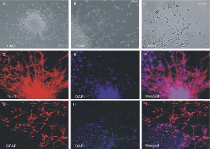
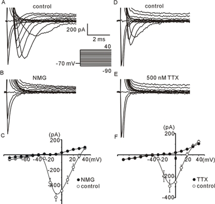
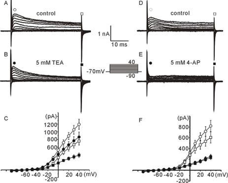
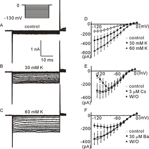
The profile of membrane currents was investigated in differentiated neuronal cells derived from human neural stem cells (hNSCs) that were obtained from aborted fetal cortex. Whole-cell voltage clamp recording revealed at least 4 different currents: a tetrodotoxin (TTX)-sensitive Na(+) current, a hyperpolarization-activated inward current, and A-type and delayed rectifier-type K(+) outward currents. Both types of K(+) outward currents were blocked by either 5 mM tetraethylammonium (TEA) or 5 mM 4-aminopyridine (4-AP). The hyperpolarization-activated current resembled the classical K(+) inward current in that it exhibited a voltage-dependent block in the presence of external Ba(2+) (30microM) or Cs(+) (3microM). However, the reversal potentials did not match well with the predicted K(+) equilibrium potentials, suggesting that it was not a classical K(+) inward rectifier current. The other Na(+) inward current resembled the classical Na(+) current observed in pharmacological studies. The expression of these channels may contribute to generation and repolarization of action potential and might be regarded as functional markers for hNSCs-derived neurons.
Keywords: A-type; Human neural stem cells; TTX-sensitive Na+ current; delayed rectifier; hyperpolarization-activated inward current.
Figures
Similar articles
-
Membrane currents in visually identified motoneurones of neonatal rat spinal cord.J Physiol. 1990 Apr;423:27-46. doi: 10.1113/jphysiol.1990.sp018009. J Physiol. 1990. PMID: 2388151 Free PMC article.
-
Potassium currents in adult rat intracardiac neurones.J Physiol. 1995 Jul 1;486 ( Pt 1)(Pt 1):15-31. doi: 10.1113/jphysiol.1995.sp020787. J Physiol. 1995. PMID: 7562632 Free PMC article.
-
Voltage-activated membrane currents in rat cerebellar granule neurones.J Physiol. 1989 Jul;414:179-99. doi: 10.1113/jphysiol.1989.sp017683. J Physiol. 1989. PMID: 2558168 Free PMC article.
-
Voltage-dependent and odorant-regulated currents in isolated olfactory receptor neurons of the channel catfish.J Gen Physiol. 1992 Apr;99(4):505-29. doi: 10.1085/jgp.99.4.505. J Gen Physiol. 1992. PMID: 1597676 Free PMC article.
-
Voltage-dependent conductances of solitary ganglion cells dissociated from the rat retina.J Physiol. 1987 Apr;385:361-91. doi: 10.1113/jphysiol.1987.sp016497. J Physiol. 1987. PMID: 2443669 Free PMC article. Review.
Cited by
-
Developmental HCN channelopathy results in decreased neural progenitor proliferation and microcephaly in mice.Proc Natl Acad Sci U S A. 2021 Aug 31;118(35):e2009393118. doi: 10.1073/pnas.2009393118. Proc Natl Acad Sci U S A. 2021. PMID: 34429357 Free PMC article.
-
Gut-derived factors promote neurogenesis of CNS-neural stem cells and nudge their differentiation to an enteric-like neuronal phenotype.Am J Physiol Gastrointest Liver Physiol. 2011 Oct;301(4):G644-55. doi: 10.1152/ajpgi.00123.2011. Epub 2011 Aug 4. Am J Physiol Gastrointest Liver Physiol. 2011. PMID: 21817062 Free PMC article.
-
Functional ion channels in stem cells.World J Stem Cells. 2011 Mar 26;3(3):19-24. doi: 10.4252/wjsc.v3.i3.19. World J Stem Cells. 2011. PMID: 21607133 Free PMC article.
References
-
- Akesson E, Piao JH, Samuelsson EB, Holmberg L, Kjaeldgaard A, Falci S, Sundstrom E, Seiger A. Long-term culture and neuronal survival after intraspinal transplantation of human spinal cordderived neurospheres. Physiol Behav. 2007;92:60–66. - PubMed
-
- Artiani L, Bettiol E, Stillitano F, Mugelli A, Cerbai E, Jaconi ME. Developmental changes in cardiomyocytes differentiated from human embryonic stem cells: a molecular and electrophysiological approach. Stem Cells. 2007;25:1136–1144. - PubMed
-
- Balasubramaniyan V, de Haas AH, Bakels R, Koper A, Boddeke HW, Copray JC. Functionally deficient neuronal differentiation of mouse embryonic neural stem cells in vitro. Neurosci Res. 2004;49:261–265. - PubMed
-
- Buzańska L, Jurga M, Domańska-Janik K. Neuronal differentiation of human umbilical cord blood neural stem-like cell line. Neurodegener Dis. 2006;3:19–26. - PubMed
