

Diversity of ion channels in human bone marrow mesenchymal stem cells from amyotrophic lateral sclerosis patients
- PMID: 19967076
- PMCID: PMC2788656
- DOI: 10.4196/kjpp.2008.12.6.337
Diversity of ion channels in human bone marrow mesenchymal stem cells from amyotrophic lateral sclerosis patients
Abstract
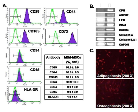
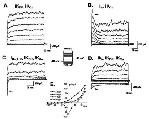
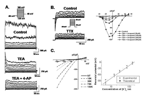
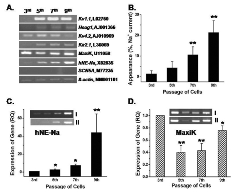
Human bone marrow mesenchymal stem cells (hBM-MSCs) represent a potentially valuable cell type for clinical therapeutic applications. The present study was designed to evaluate the effect of long-term culturing (up to 10(th) passages) of hBM-MSCs from eight individual amyotrophic lateral sclerosis (ALS) patients, focusing on functional ion channels. All hBM-MSCs contain several MSCs markers with no significant differences, whereas the distribution of functional ion channels was shown to be different between cells. Four types of K(+) currents, including noise-like Ca(+2)-activated K(+) current (IK(Ca)), a transient outward K(+) current (I(to)), a delayed rectifier K(+) current (IK(DR)), and an inward-rectifier K(+) current (K(ir)) were heterogeneously present in these cells, and a TTX-sensitive Na(+) current (I(Na,TTX)) was also recorded. In the RT-PCR analysis, Kv1.1, heag1, Kv4.2, Kir2.1, MaxiK, and hNE-Na were detected. In particular, I(Na,TTX) showed a significant passage-dependent increase. This is the first report showing that functional ion channel profiling depend on the cellular passage of hBM-MSCs.
Keywords: Bone marrow; Functional ion channels; Passage-dependency; Stem cells; Tetrodotoxin-sensitive Na+ current.
Figures
Similar articles
-
Functional expression of ion channels in mesenchymal stem cells derived from umbilical cord vein.Stem Cells. 2007 Aug;25(8):2044-52. doi: 10.1634/stemcells.2006-0735. Epub 2007 May 24. Stem Cells. 2007. PMID: 17525238
-
Characterization of ionic currents in human mesenchymal stem cells from bone marrow.Stem Cells. 2005 Mar;23(3):371-82. doi: 10.1634/stemcells.2004-0213. Stem Cells. 2005. PMID: 15749932
-
Electrophysiological properties of human adipose tissue-derived stem cells.Am J Physiol Cell Physiol. 2007 Nov;293(5):C1539-50. doi: 10.1152/ajpcell.00089.2007. Epub 2007 Aug 8. Am J Physiol Cell Physiol. 2007. PMID: 17687001
-
Ion channels in mesenchymal stem cells from rat bone marrow.Stem Cells. 2006 Jun;24(6):1519-28. doi: 10.1634/stemcells.2005-0307. Epub 2006 Feb 16. Stem Cells. 2006. PMID: 16484345
-
Regulation of cell proliferation of human induced pluripotent stem cell-derived mesenchymal stem cells via ether-à-go-go 1 (hEAG1) potassium channel.Am J Physiol Cell Physiol. 2012 Jul 15;303(2):C115-25. doi: 10.1152/ajpcell.00326.2011. Epub 2012 Feb 22. Am J Physiol Cell Physiol. 2012. PMID: 22357737
Cited by
-
Identification and functional characterization of ion channels in CD34(+) hematopoietic stem cells from human peripheral blood.Mol Cells. 2011 Aug;32(2):181-8. doi: 10.1007/s10059-011-0068-9. Epub 2011 Jun 1. Mol Cells. 2011. PMID: 21638203 Free PMC article.
References
-
- Balana B, Nicoletti C, Zahanich I, Graf EM, Christ T, Boxberger S, Ravens U. 5-Azacytidine induces changes in electrophysiological properties of human mesenchymal stem cells. Cell Res. 2006;16:949–960. - PubMed
-
- Banfi A, Muraglia A, Dozin B, Mastrogiacomo M, Cancedda R, Quarto R. Proliferation kinetics and differentiation potential of ex vivo expanded human bone marrow stromal cells:Implications for their use in cell therapy. Exp Hematol. 2000;28:707–715. - PubMed
-
- Baxter MA, Wynn RF, Jowitt SN, Wraith JE, Fairbairn LJ, Bellantuono I. Study of telomere length reveals rapid aging of human marrow stromal cells following in vitro expansion. Stem Cells. 2004;22:675–682. - PubMed
-
- Biagiotti T, D'Amico M, Marzi I, Di Gennaro P, Arcangeli A, Wanke E, Olivotto M. Cell renewing in neuroblastoma: electrophysiological and immunocytochemical characterization of stem cells and derivatives. Stem Cells. 2006;24:443–453. - PubMed
LinkOut - more resources
Full Text Sources
Miscellaneous
