

Fibrin network structure and clot mechanical properties are altered by incorporation of erythrocytes
- PMID: 19967148
- PMCID: PMC2840711
- DOI: 10.1160/TH09-03-0199
Fibrin network structure and clot mechanical properties are altered by incorporation of erythrocytes
Abstract
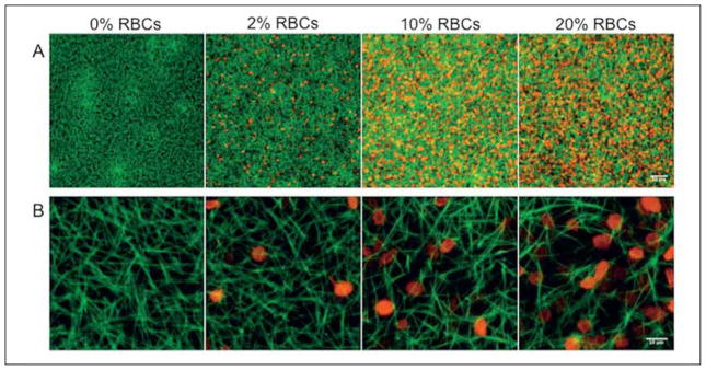
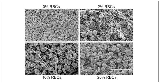
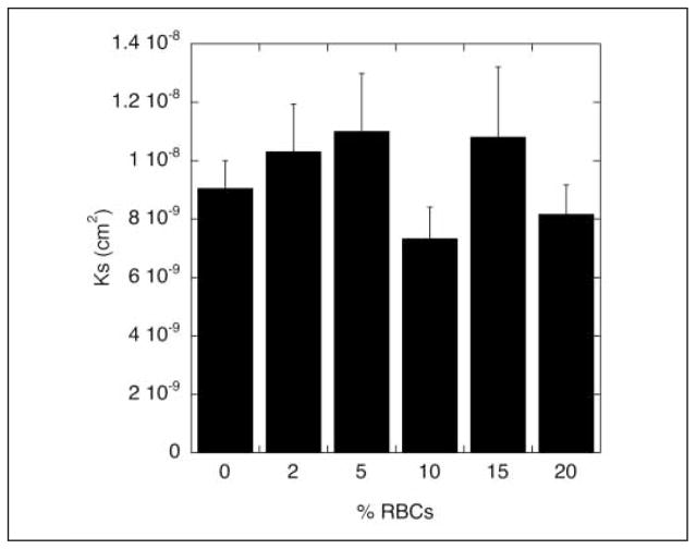
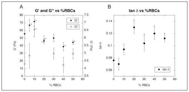
Although many in vitro fibrin studies are performed with plasma, in vivo clots and thrombi contain erythrocytes, or red blood cells (RBCs). To determine the effects of RBCs on fibrin clot structure and mechanical properties, we compared plasma clots without RBCs to those prepared with low (2 vol%), intermediate (5-10 vol%), or high (> or =20 vol%) numbers of RBCs. By confocal microscopy, we found that low RBC concentrations had little effect on clot structure. Intermediate RBC concentrations caused heterogeneity in the fiber network with pockets of densely packed fibers alongside regions with few fibers. With high levels of RBCs, fibers arranged more uniformly but loosely around the cells. Scanning electron micrographs demonstrated an uneven distribution of RBCs throughout the clot and a significant increase in fiber diameter upon RBC incorporation. While permeability was not affected by RBC addition, at 20% or higher RBCs, the ratio of viscous modulus (G'') to elastic modulus (G') increased significantly over that of a clot without any RBCs. RBCs triggered variability in the fibrin network structure, individual fiber characteristics, and overall clot viscoelasticity compared to the absence of cells. These results are important for understanding in vivo clots and thrombi.
Figures
Similar articles
-
Sensing adhesion forces between erythrocytes and γ' fibrinogen, modulating fibrin clot architecture and function.Nanomedicine. 2018 Apr;14(3):909-918. doi: 10.1016/j.nano.2018.01.006. Epub 2018 Feb 2. Nanomedicine. 2018. PMID: 29410160
-
The spatial dynamics of fibrin clot dissolution catalyzed by erythrocyte-bound vs. free fibrinolytics.J Thromb Haemost. 2010 May;8(5):1066-74. doi: 10.1111/j.1538-7836.2010.03802.x. Epub 2010 Feb 9. J Thromb Haemost. 2010. PMID: 20149071 Free PMC article.
-
Fibrin film on clots is increased by hematocrit but reduced by inflammation: implications for platelets and fibrinolysis.J Thromb Haemost. 2025 Apr;23(4):1247-1259. doi: 10.1016/j.jtha.2024.12.023. Epub 2025 Jan 2. J Thromb Haemost. 2025. PMID: 39755330
-
Fibrin mechanical properties and their structural origins.Matrix Biol. 2017 Jul;60-61:110-123. doi: 10.1016/j.matbio.2016.08.003. Epub 2016 Aug 20. Matrix Biol. 2017. PMID: 27553509 Free PMC article. Review.
-
Structure of fibrin: impact on clot stability.J Thromb Haemost. 2007 Jul;5 Suppl 1:116-24. doi: 10.1111/j.1538-7836.2007.02504.x. J Thromb Haemost. 2007. PMID: 17635717 Review.
Cited by
-
Red Cell Distribution Width Has a Predictable Value for Differentiation of Provoked and Unprovoked Venous Thromboembolism.Indian J Hematol Blood Transfus. 2016 Dec;32(4):481-487. doi: 10.1007/s12288-015-0626-y. Epub 2015 Dec 12. Indian J Hematol Blood Transfus. 2016. PMID: 27812260 Free PMC article.
-
Does prior administration of rtPA influence acute ischemic stroke clot composition? Findings from the analysis of clots retrieved with mechanical thrombectomy from the RESTORE registry.J Neurol. 2022 Apr;269(4):1913-1920. doi: 10.1007/s00415-021-10758-5. Epub 2021 Aug 20. J Neurol. 2022. PMID: 34415423 Free PMC article.
-
RBCs regulate platelet function and hemostasis under shear conditions through biophysical and biochemical means.Blood. 2024 Oct 3;144(14):1521-1531. doi: 10.1182/blood.2024023887. Blood. 2024. PMID: 38985835
-
The interplay between tissue plasminogen activator domains and fibrin structures in the regulation of fibrinolysis: kinetic and microscopic studies.Blood. 2011 Jan 13;117(2):661-8. doi: 10.1182/blood-2010-06-290338. Epub 2010 Oct 21. Blood. 2011. PMID: 20966169 Free PMC article.
-
Erythrocyte oxidative stress and thrombosis.Expert Rev Mol Med. 2022 Aug 26;24:e31. doi: 10.1017/erm.2022.25. Expert Rev Mol Med. 2022. PMID: 36017709 Free PMC article. Review.
References
-
- Zannad F, Stoltz JF. Blood rheology in arterial hypertension. J Hypertens Suppl. 1992;10:S69–78. - PubMed
-
- Schmid-Schonbein H, Wells R, Goldstone J. Influence of deformability of human red cells upon blood viscosity. Circ Res. 1969;25:131–143. - PubMed
-
- Landolfi R, Rocca B, Patrono C. Bleeding and thrombosis in myeloproliferative disorders: mechanisms and treatment. Crit Rev Oncol Hematol. 1995;20:203–222. - PubMed
-
- Goldsmith HL. Red cell motions and wall interactions in tube flow. Fed Proc. 1971;30:1578–1590. - PubMed
