. 2009 Dec;102(6):1183-93.
doi: 10.1160/TH09-04-0273.

Anticoagulant activity of a sulfated galactan: serpin-independent effect and specific interaction with factor Xa

Affiliations

Anticoagulant activity of a sulfated galactan: serpin-independent effect and specific interaction with factor Xa

Bianca F Glauser et al. Thromb Haemost. 2009 Dec.

Abstract

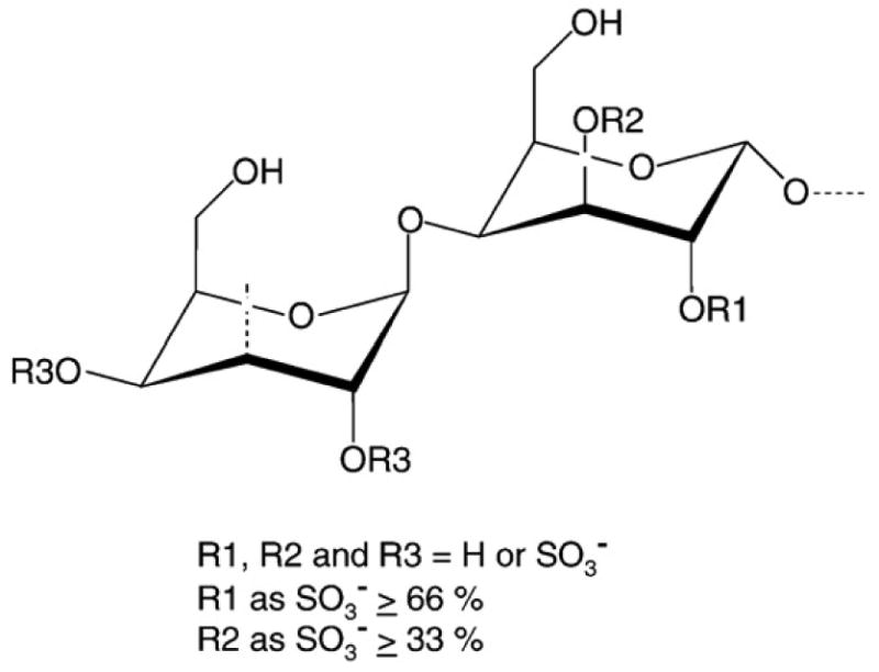
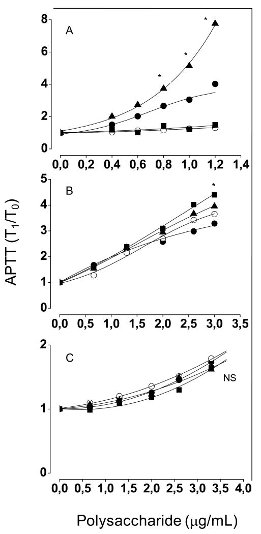
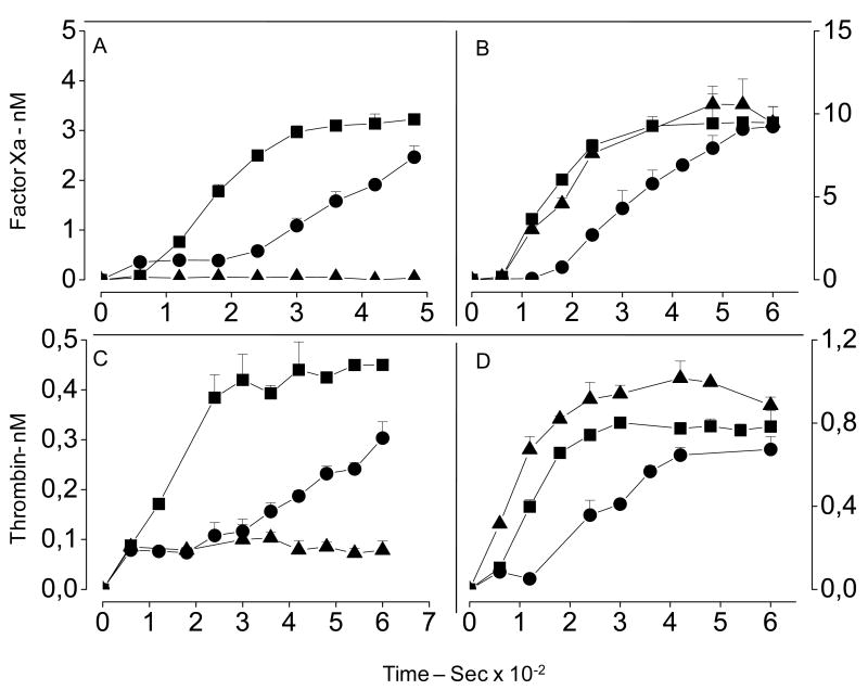
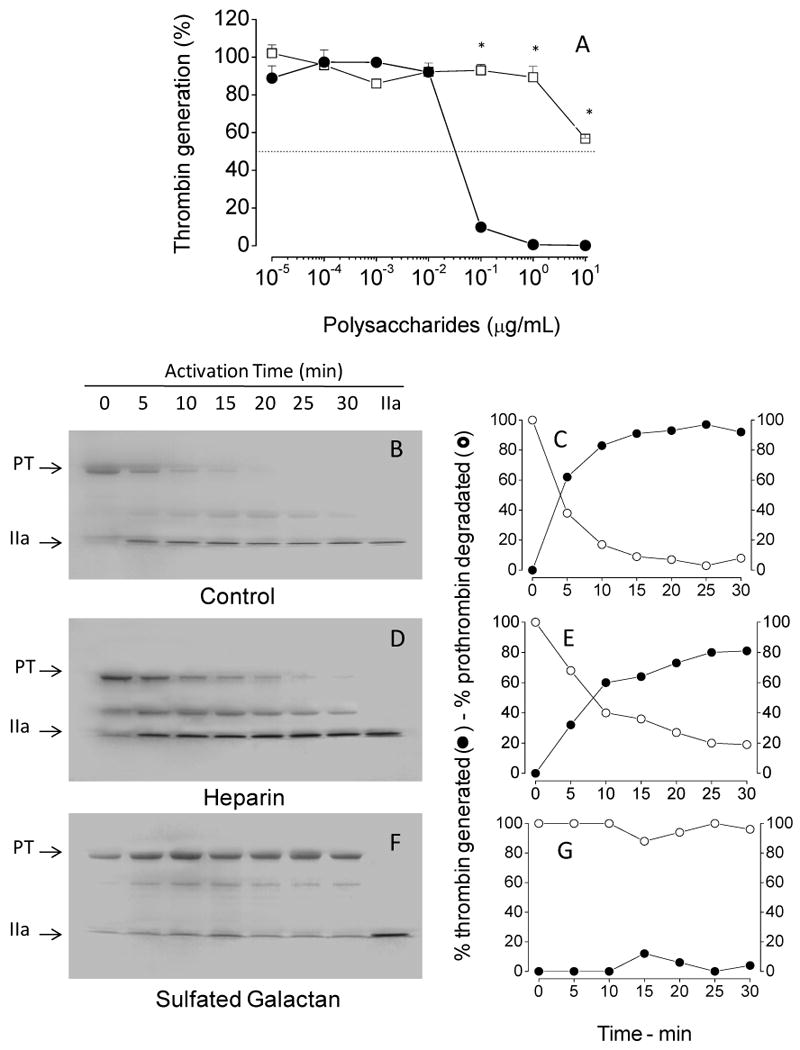
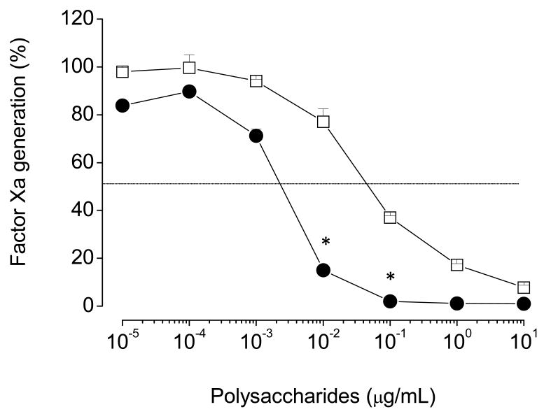
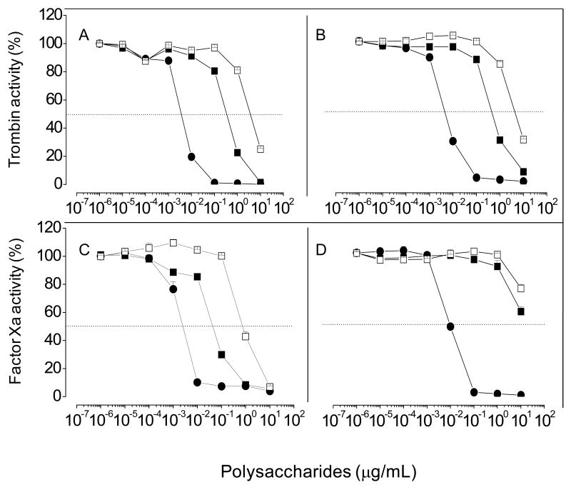
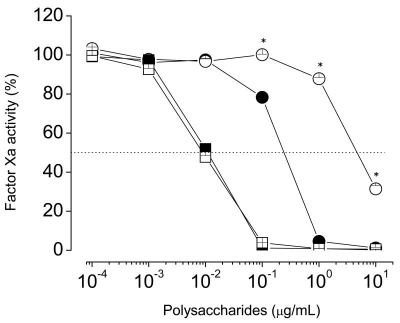
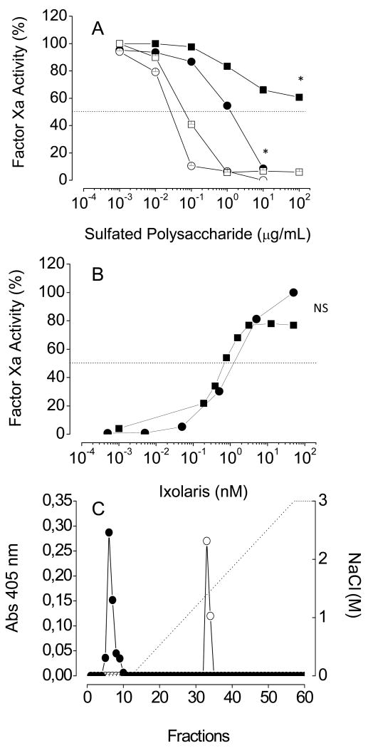
An algal sulfated galactan has high anticoagulant and antithrombotic activities. Its serpin-dependent anticoagulant action is due to promoting thrombin and factor (F)Xa inhibition by antithrombin and heparin cofactor II. Here, we evaluated the anticoagulant effect of the algal sulfated galactan using serpin-free plasma. In contrast to heparin, the sulfated galactan is still able to prolong coagulation time and delay thrombin and FXa generation in serpin-free plasma. We further investigated this effect using purified blood coagulation proteins, discovering that sulfated galactan inhibits the intrinsic tenase and prothrombinase complexes, which are critical for FXa and thrombin generation, respectively. We also investigated the mechanism by which sulfated galactan promotes FXa inhibition by antithrombin using specific recombinant mutants of the protease. We show that sulfated galactan interacts with the heparin-binding exosite of FXa and Arg-236 and Lys-240 of this site are critical residues for this interaction, as observed for heparin. Thus, sulfated galactan and heparin have similar high-affinity and specificity for interaction with FXa, though they have differences in their chemical structures. Similar to heparin, the ability of sulfated galactan to potentiate FXa inhibition by antithrombin is calcium-dependent. However, in contrast to heparin, this effect is not entirely dependent on the conformation of the gamma-carboxyglutamic acid-rich domain of the protease. In conclusion, sulfated galactan and heparin have some similar effects on blood coagulation, but also differ significantly at the molecular level. This sulfated galactan opens new perspective for the development of antithrombotic drugs.


Figures

Figure 1
Figure 1. Structure of sulfated galactan from the marine alga B. occidentalis
This polysaccharide has a repeating structure (-4-α-D-Galp-1→3-β-D-Galp-1→), with a variable sulfation pattern. Approximately one third of the total α-units are 2,3-di-sulfated, and another one-third are 2-sulfated (Farias et al. 2000).
Figure 2
Figure 2. Anticoagulant activity of heparin (A), high- (B) and low- (C) molecular-weight sulfated galactans based on APTT, performed with normal human plasma (●), antithrombin- (■), heparin cofactor II- (▲) or both serpins-free plasmas (○)
A mixture of 100 μL of normal or serpin-free human plasma and increasing concentrations of sulfated polysaccharide were incubated with 100 μL of APTT reagent. After 2 min of incubation at 37°C, 100 μL of 25 mM CaCl2 was added to the mixtures, and the clotting time was recorded. The results are expressed as ratios of clotting time in the presence (T1) and in the absence (T0) of sulfated polysaccharide. The panels show mean±SD, n=3. (A) *p < 0,05 for ▲, ■ or ○ vs. ●; (B) *p < 0,05 for ■ vs. ●; (C) NS, not significant (p > 0,05).
Figure 3
Figure 3. Factor Xa (A, and B) and thrombin (C and D) generation tests in normal (A and C) and serpin-free (B and D) plasmas
Defibrinated plasmas (50 μL) were incubated in the absence (■) or in the presence of 0.2 μg/mL of heparin (▲) or sulfated galactan (●) with 50 μL of TS/PEG buffer and 10 μL of Cephalin reagent. After incubation for 2 min at room temperature, the protease generation reaction was started by addition of 100 μL 12.5 mM CaCl2, and aliquots of 10 μL were removed each 15 s into microplate wells containing 40 μL TS/PEG buffer + 50 mM EDTA. The amounts of factor Xa or thrombin generated were determined using the chromogenic substrates S-2765 or S-2238, respectively. Substrate hydrolysis was detected using a Thermomax Microplate Reader. Reactions were recorded continuously at 405 nm for 15 min at 37°C. The panels show mean±SD, n=3.
Figure 4
Figure 4. Effect of sulfated polysaccharides on thrombin generation by the prothrombinase complex
(A) Assays based on protease activity: Increasing concentrations of sulfated galactan (●) or heparin (□) were incubated with 10 pM plasma-derived factor Xa, 1 nM factor Va and 20 μM PC/PS vesicles in TS/PEG buffer containing 10 mM CaCl2, final volume of 100 μL, for 2 min at 37°C. The prothrombinase complex was then activated by the addition of 0.5 μM human prothrombin, and after 4 min, aliquots of 10 μL were removed for the assay of thrombin activity using the chromogenic substrate S-2238, as described in the legend of Fig. 3. The panel shows mean±SD, n=3. (B-G) Assays based on SDS/PAGE: The effect of sulfated polysaccharides on prothrombinase complex was analyzed on 12% SDS-PAGE. The incubation mixtures contain: 1 nM plasma-derived factor Xa, 3 nM factor Va, 20 μM phospholipids and 10 μg/mL sulfated polysaccharide in TS/PEG buffer containing 10 mM CaCl2, final volume 500 μL. After incubation for 10 min at 37°C, the activation reaction was started by addition of 0.5 μM prothrombin. Aliquots from each reaction mixture were removed at the time point indicated in the panels and immediately quenched in SDS-PAGE loading buffer. The gel was stained with Coomassie blue and band intensities for prothrombin and thrombin were monitored by densitometric analysis. PT, prothrombin and IIa, thrombin. *p < 0,05 for □ vs. ●.
Figure 5
Figure 5. Effect of sulfated galactan (●) and heparin (□) on generation of factor Xa by the intrinsic tenase complex
Increasing concentrations of sulfated polysaccharide were incubated with a mixture containing 200 pM factor IXa, 1 unit/mL factor VIIIa and 20 μM PC/PS vesicles, in TS/PEG buffer containing 10 mM CaCl2, final volume of 100 μL, for 2 min at 37°C. The tenase complex was activated by the addition of 50 nM factor X, and 6 min later, aliquots of 25 μL were removed into microplate wells containing 25 μl TS/PEG buffer + 50 mM EDTA. The amount of factor Xa formed was determined after the addition of 50 μL of 100 μM chromogenic substrate S-2765, as described in the legend of Fig. 3. The panels show mean±SD, n=3. *p < 0,05 for □ vs. ●.
Figure 6
Figure 6. Effect of Ca2 on heparin- or sulfated galactan-catalyzed inactivation of thrombin (A, B) or factor Xa (C, D) by antithrombin
Thrombin (0.5 nM) or factor Xa (0.5 nM) inactivation by antithrombin (5 nM) was monitored at increasing concentrations of heparin (●), high- (■) or low- (□) molecular-weigth sulfated galactan in TS/PEG buffer, containing either 10 mM CaCl2 (A and C) or 20 mM EDTA (B and D). After 60 s, the remaining thrombin or factor Xa activity was determined by the addition of S-2238 or S-2765, respectively, as described in the legend of Fig. 3. The panels show mean±SD, n=3.
Figure 7
Figure 7. Effect of Ca2 on heparin- or sulfated galactan-catalyzed inactivation of GD-factor Xa by antithrombin
Inactivation of recombinant GD-factor Xa (0.5 nM) by antithrombin (5 nM) was monitored at increasing concentrations of heparin (squares) and sulfated galactan (circles) in TS/PEG buffer, containing either 10 mM CaCl2 (closed symbols) or 20 mM EDTA (open symbols). The total volume of reaction mixture was 100 μL. After 60 s, the remaining factor Xa activity was determined by the addition of S-2238 or S-2765 susbtrate (25 μL of a 200 μM solution), and substrate hydrolysis was monitored as described in the legend of Fig. 3. *p < 0,05 for ○ vs.●.
Figure 8
Figure 8. Effect of Ixolaris on antithrombin-mediated factor Xa inactivation by heparin or sulfated galactan
(A) Dependence on the concentration of sulfated polysaccharide: Increasing concentrations of heparin (squares) or sulfated galactan (circles) were incubated with 0.5 nM plasma-derived factor Xa and 5 nM antithrombin, in the absence (open symbols) or in the presence (closed symbols) of 10 nM Ixolaris, in TS/PEG buffer containing 10 mM CaCl2. The total volume of reaction mixtures was 100 μL. After 60 s, the remaining factor Xa activity activity was determined using S-2765 substrate. (B) Dependence on the concentration of Ixolaris: The assays were as described for Panel A, except that increasing concentration of Ixolaris and fixed concentrations of heparin (1 μg/ml) or sulfated galactan (10 μg/ml) were used. (C) Sulfated galactan-affinity chromatography: One mL of plasma-derived factor Xa (100 nM), before (○) or after (●) incubation with 200 nM Ixolaris in TS/PEG buffer, containing 50 mM CaCl2 was applied to a sulfated galactan-Sepharose column (1 mL), connected to a HPLC system. The column was washed with 5 mL of the TS/PEG buffer and eluted with a linear gradient of 0.15 - 3 M NaCl, at a flow rate of 0.5 mL/min. Fractions of 0.5 mL were collected and checked by factor Xa activity using S-2765 substrate. *p < 0,05 for ■ vs. □ or ○ vs. ●; NS, not significant (p > 0,05).
Figure 9
Figure 9. Interaction of GD-factor Xa with sulfated galactan. Concentration-dependence of high- (A) or low- (B) molecular-weight sulfated galactan to catalyze antithrombin-mediated inactivation of GD-factor Xa
Increasing concentrations of the sulfated galactan were incubated with 5 nM antithrombin and 0.5 nM of wild or mutant Gla domainless factor Xa in TS/PEG buffer, final volume 100 μL, for 5 min at 37°C. Residual protease activity was determined by the addition of 100 μM of the chromogenic substrate S-2765, as described in the legend of Fig. 3. The symbols are as follows: wild-type (■), R125A (□), R93A (○), K96A (+), R165A (▲), K236A (△), K169A (●) and R240A (◆). (C) Sulfated galactan-affinity chromatography: Wild (■), R93A (○) or K236A (△) mutants of Gla domainless factor Xa (100 nM of each) were applied on a 1 mL sulfated galactan-Sepharose column, pre-equilibrated in TS/PEG buffer. The column was washed with 5 mL of the same solution at a flow rate of 0.5 mL/min, and then eluted with a 0.15 - 3 M NaCl gradiente. Fractions of 0.5 mL were collected and checked for factor Xa activity, as described in the legend of Fig. 3. Mutant R240A was not retained on the column, while mutants R96A, R125A and R169A were eluted from the column at 0.75 M NaCl, as the wild protease and the R93A mutant. (A) *p < 0,05 for ◆, △, ▲ vs ■ (B) *p < 0,05 for ◆, △ vs ■.

Similar articles

Cited by

References

    1. Fareed J, Hoppensteadt DA. Heparins in the new millennium: Will unfractionated heparin survive? Semin Thromb Haemost. 2000;26:87–88. - PubMed
    1. Béguin S, Lindhout T, Hemker HC. The mode of action of heparin in plasma. Thromb Haemost. 1988;60:457–62. - PubMed
    1. Blossom DB, et al. Outbreak of Adverse Reactions Associated with Contaminated Heparin. N Engl J Med. 2008;359:2674–84. - PMC - PubMed
    1. Mourão PAS, Pereira MS. Searching for alternatives to heparin. Trends Cardiovasc Med. 1999;9:225–32. - PubMed
    1. Farias WRL, Valente AP, Pereira MS, Mourão PAS. Structure and anticoagulant activity of sulfated galactans - Isolation of a unique sulfated galactan from the red algae Botryocladia occidentalis and comparison of its anticoagulant action with that of sulfated galactans from invertebrates. J Biol Chem. 2000;275:29299–307. - PubMed

Publication types

MeSH terms