

Review
Illuminating rationale and uses for light therapy
Affiliations
- PMID: 19968050
- PMCID: PMC2670336
Item in Clipboard
Review
Illuminating rationale and uses for light therapy
J Clin Sleep Med.
.
Abstract
Light therapy is increasingly applied in a variety of sleep medicine and psychiatric conditions including circadian rhythm sleep disorders, seasonal affective disorder, and dementia. This article reviews the neural underpinnings of circadian neurobiology crucial for understanding the influence of light therapy on brain function, common mood and sleep disorders in which light therapy may be effectively used, and applications of light therapy in clinical practice.
Figures
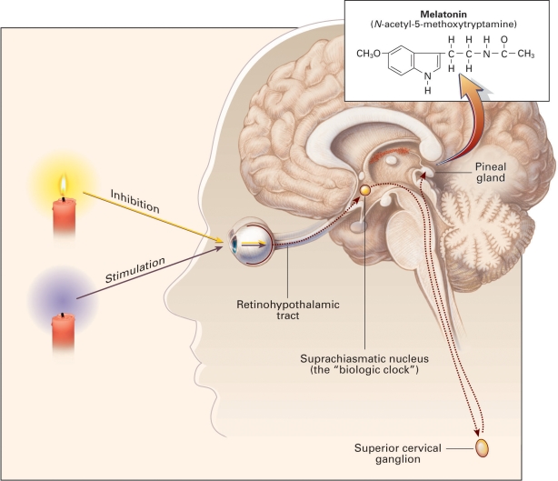
Physiology of Melatonin Secretion. Melatonin (inset) is produced in the pineal gland. The production and secretion of melatonin are mediated largely by postganglionic retinal nerve fibers that pass through the retinohypothalamic tract to the suprachiasmatic nucleus, then to the superior cervical ganglion, and finally to the pineal gland. This neuronal system is activated by darkness and suppressed by light. The activation of α1- and β1-adrenergic receptors in the pineal gland raises cyclic AMP and calcium concentrations and activates arylalkylamine N-acetyltranferanse, initiating the synthesis and release of melatonin. The daily rhythm of melatonin secretion is also controlled by an endogenous, free-running pacemaker located in the suprachiasmatic nucleus. Reproduced from Brzezinski A. Melatonin in humans. N Eng J Med 1997; 336:186-195 (with kind permission from The Publishing Division of the Massachusetts Medical Society, Waltham, Massachusetts).
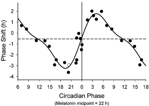
The PRC to the bright light stimulus using melatonin midpoints as the circadian phase marker. Phase advances (positive values) and delays (negative values) are plotted against the timing of the centre of the light exposure relative to the melatonin midpoint on the pre-stimulus constant routine (defined to be 22 h), with the core body temperature minimum assumed to occur 2 h later at 0 h. Data points from circadian phases 6-18 are double plotted. The filled circles represent data from plasma melatonin, and the open circle represents data from salivary melatonin in subject 18K8 from whom blood samples were not acquired. The solid curve is a dual harmonic function fitted through all of the data points. The horizontal dashed line represents the anticipated 0.54 h average delay drift of the pacemaker between the pre- and post-stimulus phase assessments. Reproduced from: Khalsa SBS, Jewett ME, Cajochen C, Czeisler CA. A phase response curve to single bright light pulses in human subjects. J Physiol 2003; 549: 945–952 (with kind permission of Wiley-Blackwell, Oxford, UK).
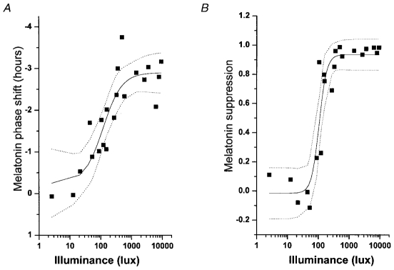
Illuminance-response curve of the human circadian pacemaker. The shift in the phase of the melatonin rhythm (A), as assessed on the day following exposure to a 6·5 h experimental light stimulus, has been fitted with a four parameter logistic model using a non-linear least squares analysis. Acute suppression of plasma melatonin (B) during the light exposure also has been fitted with a four parameter logistic model using a non-linear least squares analysis. The logistic models predict an inflection point of the curve (i.e. the sensitivity of the system) at ≈120 lx. Saturation of the phase-shift response is predicted to occur with ≈550 lx and saturation of the melatonin-suppression response is predicted to occur with ≈200 lx. Individual subjects are represented by ▪ the model by the continuous line, and the 95 % confidence intervals by the dotted lines. Reproduced from: Zeitzer JM, Dijk DJ, Kronauer RE, Brown EN, Czeisler CA. Sensitivity of the human circadian pacemaker to nocturnal light: melatonin phase resetting and suppression. J Physiol 2000; 526: 695-702 (with kind permission of Wiley-Blackwell, Oxford, UK).
References
-
- Sulzman FM, Ellman D, Fuller CA, Moore-Ede MC, Wassmer G. Neurospora circadian rhythms in space: a reexamination of the endogenous-exogenous question. Science. 1984;225:232–4. - PubMed
-
- Ralph MR, Foster RG, Davis FC, Menaker M. Transplanted suprachiasmatic nucleus determines circadian period. Science. 1990;247:975–8. - PubMed
-
- Moore RY, Eichler VB. Loss of a circadian adrenal corticosterone rhythm following suprachiasmatic lesions in the rat. Brain Res. 1972;42:201–6. - PubMed
-
- Okamura H, Miyake S, Sumi Y, et al. Photic induction of mPer1 and mPer2 in Cry-deficient mice lacking a biological clock. Science. 1999;286:2531–4. - PubMed
Publication types
MeSH terms
LinkOut - more resources
Full Text Sources
Other Literature Sources
Medical