. 2010 Feb 9;426(2):171-81.
doi: 10.1042/BJ20091439.

Oct-4 controls cell-cycle progression of embryonic stem cells

Affiliations

Oct-4 controls cell-cycle progression of embryonic stem cells

Jungwoon Lee et al. Biochem J. .

Abstract

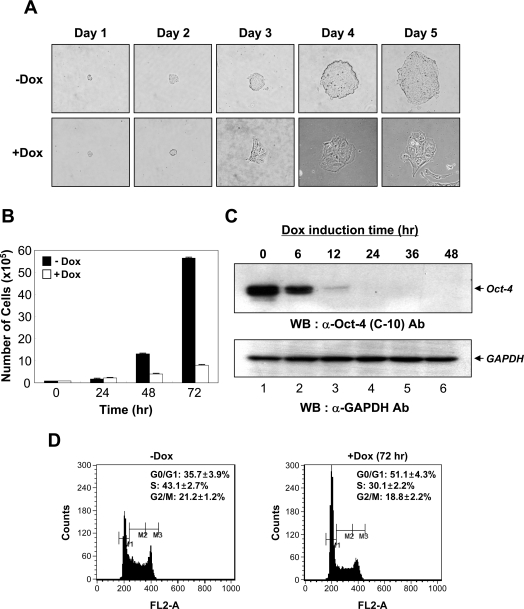
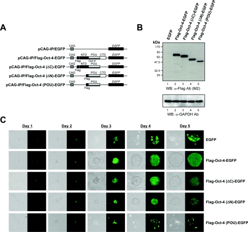
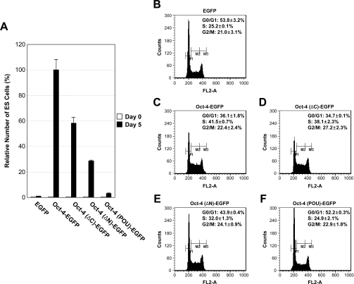
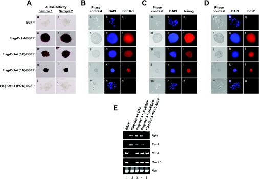
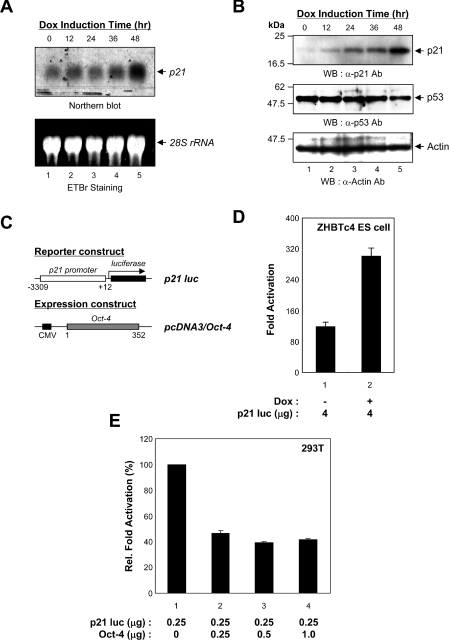
Mouse and human ES (embryonic stem) cells display unusual proliferative properties and can produce pluripotent stem cells indefinitely. Both processes might be important for maintaining the 'stemness' of ES cells; however, little is known about how the cell-cycle fate is regulated in ES cells. Oct-4, a master switch of pluripotency, plays an important role in maintaining the pluripotent state of ES cells and may prevent the expression of genes activated during differentiation. Using ZHBTc4 ES cells, we have investigated the effect of Oct-4 on ES cell-cycle control, and we found that Oct-4 down-regulation in ES cells inhibits proliferation by blocking cell-cycle progression in G0/G1. Deletion analysis of the functional domains of Oct-4 indicates that the overall integrity of the Oct-4 functional domains is important for the stimulation of S-phase entry. We also show in the present study that the p21 gene is a target for Oct-4 repression. Furthermore, p21 protein levels were repressed by Oct-4 and were induced by the down-regulation of Oct-4 in ZHBTc4 ES cells. Therefore the down-regulation of p21 by Oct-4 may contribute to the maintenance of ES cell proliferation.


Figures

Figure 1
Figure 1. Growth inhibition of ES cells by the down-regulation of Oct-4
(A) Time-dependent effects of the down-regulation of Oct-4 on ES cell growth and colony morphology. ZHBTc4 ES cells (3×10³) were plated on to 0.2% gelatin-coated 35-mm dishes and treated without (labelled as −Dox) or with 1 μg/ml doxycycline (labelled as +Dox) for 5 days. Cells were monitored daily using an inverted phase-contrast microscope (IX71; Olympus). (B) Effects of Oct-4 down-regulation on the proliferation of ZHBTc4 ES cells. ZHBTc4 ES cells were seeded at 1×10⁵ cells per 0.2% gelatin-coated 60-mm dish and grown in ES cell medium supplemented with (+Dox) or without (−Dox) doxycycline. Cells were counted at 24 h intervals with a haemocytometer for a total of 72 h. Values are means±S.D. Three independent experiments were performed, all of which gave similar results. (C) Western blot analysis of Oct-4 down-regulation in ZHBTc4 ES cells following doxycycline treatment. ZHBTc4 ES cells were treated with 1 μg/ml doxycycline and were extracted 0, 6, 12, 24, 36 and 48 h after doxycycline treatment. The extracted proteins were resolved by SDS/PAGE (12% gels), transferred on to a PVDF membrane, and immunoblotted with anti-Oct-4 antibody (C-10; Santa Cruz Biotechnology) (upper panel). The expression level of GAPDH was used as an internal control (bottom panel). (D) Effects of Oct-4 down-regulation on cell-cycle progression. The DNA contents of ZHBTc4 ES cells cultured in the absence (−Dox) or presence (+Dox) of doxycycline for 72 h were measured by fluorescence with PI staining. M1, M2 and M3 represent gating for G0/G1, S and G2/M populations respectively. Values given are means±S.D. Three independent experiments gave similar results. Ab, antibody; WB, Western blot.
Figure 2
Figure 2. Different abilities of the Oct-4 deletion mutants to confer self-renewal
(A) Schematic representation of the Oct-4–EGFP fusion construct and its derivatives with deleted domains. The functional domains located within Oct-4 are indicated as NTD (N-terminal domain), POU (POU DNA-binding domain) and CTD (C-terminal domain). (B) Immunoblot analysis of the expression of Oct-4 deletion mutants in stably transfected ZHBTc4 cells. ZHBTc4 ES cells were stably transfected with pCAG-IP/EGFP (labelled as EGFP; lane 1), pCAG-IP/FLAG-Oct-4–EGFP (Flag-Oct-4-EGFP; lane 2), pCAG-IP/FLAG-Oct-4(ΔC)–EGFP [Flag-Oct-4(ΔC)-EGFP; lane 3], pCAG-IP/FLAG-Oct-4(ΔN)–EGFP [Flag-Oct-4(ΔN)-EGFP; lane 4] or pCAG-IP/FLAG-Oct-4(POU)–EGFP [Flag-Oct-4(POU)-EGFP; lane 5], and total cell lysates were fractionated by SDS/PAGE (12% gels) and visualized by Western blotting with anti-FLAG (M2; Sigma) or anti-GAPDH (V-18; Santa Cruz Biotechnology) antibodies. The position of the prestained molecular-mass marker (New England Biolabs) is indicated to the left of the gel (molecular mass in kDa). (C) Self-renewal potentials of Oct-4 deletion mutants. ZHBTc4 ES cells stably transfected with pCAG-IP/EGFP (EGFP), pCAG-IP/FLAG-Oct-4–EGFP (Flag-Oct-4-EGFP), pCAG-IP/FLAG-Oct-4(ΔC)–EGFP [Flag-Oct-4(ΔC)-EGFP], pCAG-IP/FLAG-Oct-4(ΔN)–EGFP [Flag-Oct-4(ΔN)EGFP] or pCAG-IP/FLAG-Oct-4(POU)–EGFP [Flag-Oct-4(POU)-EGFP] were cultured in the presence of doxycycline. Phase-contrast (left-hand panels, grey colour) and fluorescence views (right-hand panels, green colour) are shown. Ab, antibody; WB, Western blot.
Figure 3
Figure 3. Functional regions of Oct-4 required for stimulation of S-phase entry of ES cells
(A) Relative growth rate of ZHBTc4 ES cells expressing Oct-4 deletion mutants. Cells (3×10³) were plated on to 0.2% gelatin-coated 35-mm dishes and counted at 5 days with a haemocytometer. Relative self-renewal ability values are shown to the right, with the value obtained by Oct-4 taken as 100%. Each bar represents the average values of two experiments. Three independent experiments gave similar results. (BF) Effects of Oct-4 deletion mutants on ES cell-cycle progression. DNA contents of ZHBTc4 ES cells expressing EGFP (B), Oct-4–EGFP (C), Oct-4(ΔC)–EGFP (D), Oct-4(ΔN)–EGFP (E) or Oct-4(POU)–EGFP (F) were measured by fluorescence with PI staining. M1, M2 and M3 represent gating for G0/G1, S and G2/M populations respectively. Values shown are means±S.D. Three independent experiments gave similar results.
Figure 4
Figure 4. Characterization of ZHBTc4 ES cells expressing Oct-4 deletion mutants
(A) Expression of AP. AP activity was assessed in ZHBTc4 ES cells expressing Oct-4 deletion mutants. The expression vectors used in this experiment were the same as those described in Figure 2(A). (B) Expression of SSEA-1. The expression of SSEA-1 in ZHBTc4 ES cells expressing Oct-4 deletion mutants was analysed with an anti-SSEA-1 antibody (480; Santa Cruz Biotechnology). The expression vectors used in this experiment were the same as those described in in Figure 2(A). (C) Expression of Nanog. The expression of Nanog in ZHBTc4 ES cells expressing Oct-4 deletion mutants was analysed with an anti-Nanog antibody (ab21603; Abcam). The expression vectors used in this experiment were the same as those described in Figure 2(A). (D) Expression of Sox2. The expression of Sox2 in ZHBTc4 ES cells expressing Oct-4 deletion mutants was analysed with an anti-Sox2 antibody (Y-17; Santa Cruz Biotechnology). The expression vectors used in this experiment were the same as those described in Figure 2(A). (E) Expression of Oct-4 downstream target genes and lineage-specific markers. RT–PCR analyses of Fgf-4, Rex-1, Cdx-2 and Hand-1 mRNAs were performed in ZHBTc4 ES cells expressing Oct-4 deletion mutants. Hprt was used as a control to qualify the RT–PCR results. Following amplification, an aliquot of each product was analysed by staining the gel with ethidium bromide. The ES cell lines from which the input RNAs used in the RT reactions were derived are shown above the panel. Three independent experiments gave similar results.
Figure 5
Figure 5. Up-regulation of p21 gene expression in ZHBTc4 ES cells down-regulating Oct-4
(A) Northern blot analysis of p21 mRNA expression. ZHBTc4 ES cells were harvested for the preparation of total RNA at 0, 12, 24, 36 and 48 h following doxycycline treatment. Total RNA was fractionated on a 6% formaldehyde/1.5% agarose gel, transferred on to a nylon membrane, and probed with mouse p21 cDNA as described in the Materials and methods section (upper panel). The ethidium bromide staining of the agarose gel used for the Northern blotting is shown to demonstrate that equal amounts of total RNA were loaded into each lane (lower panel). Arrows indicate the position of migration of the respective RNAs. Three independent experiments gave similar results. (B) Western blot analysis of p21 expression. Total cell extracts were prepared from ZHBTc4 ES cells at 0, 12, 24, 36 and 48 h following doxycycline treatment. The extracts were resolved by SDS/PAGE (15% gel), transferred on to a PVDF membrane, and immunoblotted with anti-p21 (F5; Santa Cruz Biotechnology), anti-p53 (CM5; Novocastra Immunohistochemistry) or anti-actin (I-19; Santa Cruz Biotechnology) antibodies. The positions of the prestained molecular-mass markers (New England Biolabs) are indicated to the left of the gel (molecular mass in kDa). Three independent experiments gave similar results. (C) Schematic representation of the reporter plasmid and Oct-4 expression vector. The p21 luciferase reporter plasmid contains a mouse genomic DNA sequence from nucleotides −3309 to +12. The mouse p21 promoter is indicated by an open box and the luciferase gene is indicated by a solid box. The expression vector driving the production of mouse Oct-4 is also represented. (D) Transcriptional activation of the p21 promoter by down-regulation of Oct-4 in ZHBTc4 ES cells. ZHBTc4 ES cells were transfected with the p21 luciferase reporter plasmid and cultured in the absence (−Dox, lane 1) or presence (+Dox, lane 2) of doxycycline. Firefly luciferase activity was normalized with Renilla luciferase activity to correct for transfection efficiencies. Each transfection was performed at least three times independently, and values are means±S.D. Fold-induction is relative to the empty vector. (E) Transcriptional repression of the p21 promoter by Oct-4. Co-transfections of HEK-293T cells were performed with 0.25 μg of reporter plasmid and 0, 0.25, 0.5 or 1.0 μg of pcDNA3/Oct-4 expression plasmid. Individual transfected DNAs were adjusted to contain equal amounts of total DNA by the addition of the empty expression vector pcDNA3. The average relative transactivation and standard error is presented, with the relative transactivation values obtained with pcDNA3/Oct-4 taken as 100%. The values shown are taken from three independent experiments.

Similar articles

Cited by

References

    1. Niwa H. Molecular mechanism to maintain stem cell renewal of ES cells. Cell Struct. Funct. 2001;26:137–148. - PubMed
    1. Okamoto K., Okazawa H., Okuda A., Sakai M., Muramatsu M., Hamada H. A novel octamer binding transcription factor is differentially expressed in mouse embryonic cells. Cell. 1990;60:461–472. - PubMed
    1. Scholer H. R., Dressler G. R., Balling R., Rohdewohld H., Gruss P. Oct-4: a germline-specific transcription factor mapping to the mouse t-complex. EMBO J. 1990;9:2185–2195. - PMC - PubMed
    1. Rosner M. H., Vigano M. A., Ozato K., Timmons P. M., Poirier F., Rigby P. W., Staudt L. M. A POU-domain transcription factor in early stem cells and germ cells of the mammalian embryo. Nature. 1990;345:686–692. - PubMed
    1. Goto T., Adjaye J., Rodeck C. H., Monk M. Identification of genes expressed in human primordial germ cells at the time of entry of the female germ line into meiosis. Mol. Hum. Reprod. 1999;5:851–860. - PubMed

Publication types

Substances