. 2010 May;40(6):683-93.
doi: 10.1016/j.ijpara.2009.11.008. Epub 2009 Dec 5.

Characterisation of the protective immune response following subcutaneous vaccination of susceptible mice against Trichuris muris

Affiliations

Characterisation of the protective immune response following subcutaneous vaccination of susceptible mice against Trichuris muris

Helen Dixon et al. Int J Parasitol. 2010 May.

Abstract

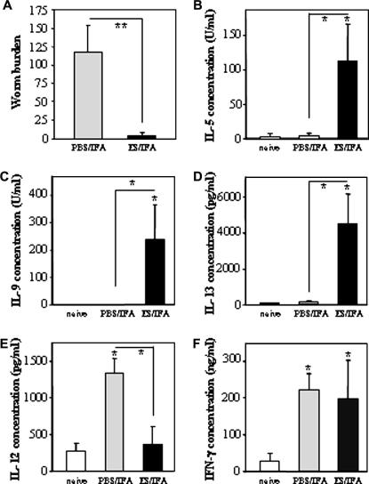
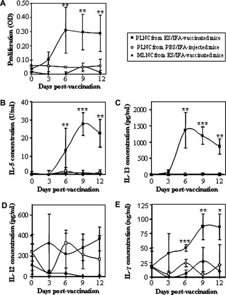
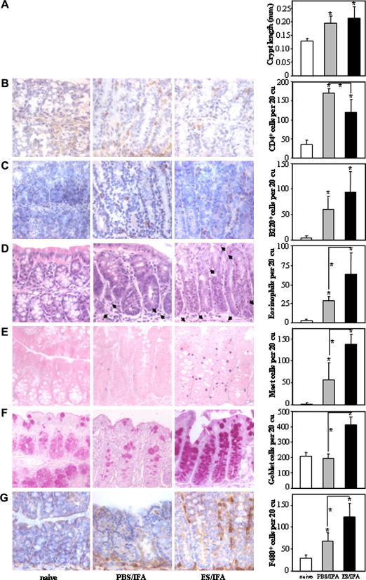
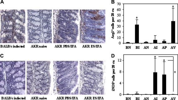
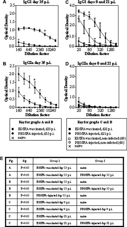
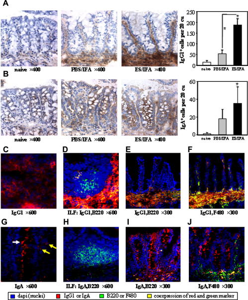
Trichuris muris is a laboratory model for the human whipworm Trichuris trichiura which infects approximately 1 billion people in tropical and sub-tropical countries. The development of a vaccine would control trichuriasis by promoting the acquisition of immunity during childhood, thereby reducing faecal egg output by the community into their environment. Resistance to T. muris, defined as expulsion of the parasite prior to patency, requires the development of a T helper 2 (Th2) response during a primary infection. To our knowledge this is the first study to describe the protective immune response in the peripheral lymph nodes (PLN), mesenteric lymph nodes (MLN) and colonic mucosa following s.c. vaccination against T. muris. Susceptible AKR mice were either vaccinated with T. muris excretory-secretory product (ES) in incomplete Freund's adjuvant (IFA) (ES/IFA) or injected with PBS in IFA (PBS/IFA) and for protection experiments were infected with embryonated infective T. muris eggs 10 days later. The ES/IFA vaccine induced the proliferation of PLN cells and their production of Th2 cytokines and the Th1-associated cytokine IFN-gamma. Following a challenge infection, the ES/IFA vaccination offered susceptible mice complete protection. While MLN-derived IFN-gamma was produced by infected mice following either ES/IFA vaccination or PBS/IFA, the protection of susceptible mice by ES/IFA was characterised by the production of MLN-derived Th2 cytokines. Goblet cell hyperplasia and the influx and alternative activation of macrophages were observed locally in the gut post-challenge infection. The rate of epithelial turnover did not appear to be increased by vaccination, suggesting that there are differences in the mechanisms of expulsion between 'natural resistance' and 'vaccinated resistance'. High levels of serum IgG1 and cell-bound IgG1 in the colon of mice protected by the ES/IFA vaccine suggest that antibody may be involved in vaccination-induced worm expulsion.


Figures

None
Graphical abstract
Fig. 1
Fig. 1
Worm burden and mesenteric lymph node cell (MLNC) cytokine profile at day 21 p.i. (A) Worm burdens and (B–F) cytokine analyses of MLNC supernatants on day 21 p.i. with Trichuris muris in naive, PBS/incomplete Freund’s adjuvant (IFA)-injected and excretory–secretory product (ES)/IFA-vaccinated AKR mice. Mice in the PBS/IFA-injected and ES/IFA-vaccinated groups were infected with approximately 150 infective embryonated T. muris eggs on day 0. MLNC supernatants were analysed for the presence of (B) IL-5, (C) IL-9, (D) IL-13, (E) IL-12p40 and (F) IFN-γ. Results are shown as means ± SD (= 5 per group). * above error bar indicates cytokine levels significantly above naive levels (< 0.05). A significant difference between infected groups is indicated by * with a line (< 0.05) or ** with a line (< 0.01). The results presented are from one of two experiments.
Fig. 2
Fig. 2
Cell proliferation and cytokine production profiles of peripheral lymph node cells (PLNC) and mesenteric lymph node cells (MLNC) following excretory–secretory product (ES)/incomplete Freund’s adjuvant (IFA) vaccination or PBS/IFA injection. (A) Cell proliferation as measured by MTT [3-(4,5-dimethylthiazol-2-yl)-2,5-diphenyl tetrazolium bromide] eluted stain assay and (B–E) cytokine analyses of in vitro ES-re-stimulated MLNC (triangles) and PLNC (squares) harvested from PBS/IFA-injected (open symbols) and ES/IFA-vaccinated (black symbols) AKR mice. Mice were vaccinated 12, 9, 6 and 3 days prior to autopsy. Results are shown as means ± SD. For all time-points = 6 per group except in the MTT assay where on day 6 = 2 for PLNC from PBS/IFA-injected AKR mice. Significant differences between the three groups are indicated by ∗< 0.05, ∗∗< 0.01, ∗∗∗< 0.001. The results presented are from one of two experiments.
Fig. 3
Fig. 3
Colonic crypt length and cell infiltration. Representative photographs and numbers of (A) colonic crypt lengths and (B) CD4+ (C) B220+ (D) eosinophil (E) mast (F) goblet and (G) F4/80+ cells in the colonic mucosa of naive and infected PBS/incomplete Freund’s adjuvant (IFA)-injected and excretory–secretory product (ES)/IFA-vaccinated AKR mice on day 21 p.i. CD4+, B220+ and F4/80+ cells appear brown in immunohistochemical (IHC) stained sections. Eosinophils have a blue nucleus and pink cytoplasm in H&E stained sections and examples are marked by an arrow. Mast cells appear blue and granular in toluidine blue while goblet cells appear pink-purple in Schiff’s stained sections. All photographs shown were taken at 400× original magnification. Graphed results are shown as mean cell numbers per 20 crypt units (cu) ± SD (= 5 per group). * above the error bar indicates that cell numbers are significantly above naive levels (< 0.05). A significant difference between infected groups is indicated by * with a line (< 0.05). The results presented are from one of two experiments.
Fig. 4
Fig. 4
Excretory–secretory product (ES)/incomplete Freund’s adjuvant (IFA) vaccination induces alternatively activated macrophages. (A and C) Representative photographs and (B and D) numbers of (A and B) Arg1+ and (C and D) iNOS+ cells in the colonic mucosa of: naive BALB/c (BN); naive AKR (AN); infected BALB/c (BI); infected AKR (AI); PBSIFA-injected and infected AKR (AP); ES/IFA-vaccinated and infected AKR (AV) mice on day 21 p.i. Arg1+ and iNOS+ cells appear brown in immunohistochemical (IHC) stained sections. All photographs were taken at 400× original magnification. Graphed results are shown as mean cell numbers per 20 crypt units (cu) ± SD (= 5 per group). (B and D) * above the error bar indicates that cell numbers are significantly above naive levels (< 0.05). (D) * with a line indicates a significant difference (< 0.05) between the infected PBS/IFA-injected and ES/IFA-vaccinated groups and between the infected non-vaccinated AKR and ES/IFA-vaccinated groups. The results presented are from one of two experiments.
Fig. 5
Fig. 5
Serum antibody responses following ES/IFA vaccination. Sera from naive and Trichuris muris-infected PBS/incomplete Freund’s adjuvant (IFA)-injected and excretory–secretory product (ES)/IFA-vaccinated AKR mice were assayed by ELISA for parasite-specific (A and C) IgG1 and (B and D) IgG2a at (A and B) day 35 p.i. and (C) IgG1 and (D) IgG2a levels are compared between day 0 and day 21 p.i. Results are shown as means ± SD (= 5 per group). Naive levels are depicted by a cross, PBS/IFA-injected levels by a circle and ES/IFA-vaccinated levels by a square. Non-infected groups are indicated by open symbols while infected groups are indicated by closed symbols. Statistical analyses were carried out from data from the (A) 1:5,120 dilution, from the (B) 1:1,280 dilution and from the (C and D) 1:80 dilution. Significant differences between groups are represented by a * and a table of significant differences between groups is presented in (E). The results presented are from one of two experiments.
Fig. 6
Fig. 6
Mucosal antibody responses following ES/IFA vaccination. (A and B) Colorimetric immunohistochemical (IHC) staining and numbers of (A) IgG1+ and (B) IgA+ (IgA+ epithelial and goblet cells not counted) leukocytes in the colonic mucosa of naive and Trichuris muris-infected PBS/incomplete Freund’s adjuvant (IFA)-injected and excretory–secretory product (ES)/IFA-vaccinated mice at day 21 p.i. IgG1+ and IgA+ cells appear brown in colorimetric IHC stained sections. Graphed results are shown as mean cell numbers per 20 crypt units (cu) ± SD (= 5 per group). * above the error bar indicates that cell number is significantly above naive levels (< 0.05). * with a line indicates a significant difference in IgG1+ cell numbers (< 0.05) between the infected groups. (C–J) Fluorescent IHC staining of colonic sections from infected ES/IFA-vaccinated mice showing (C) IgG1+ (D) ILF B220+ and IgG1+ (E) B220+ and IgG1+, (F) F4/80+ and IgG1+, (G) IgGA+ (H) ILF B220+ and IgA+ (I) B220+ and IgA+, (J) F4/80+ and IgA+ cells. (G) White arrow depicts goblet cell staining while yellow arrows depict leukocyte staining for IgA. The results presented are from one of two experiments.

Similar articles

Cited by

References

    1. Anthony R.M., Urban J.F., Jr., Alem F., Hamed H.A., Rozo C.T., Boucher J.L., van R.N., Gause W.C. Memory T(H)2 cells induce alternatively activated macrophages to mediate protection against nematode parasites. Nat. Med. 2006;12:955–960. - PMC - PubMed
    1. Artis D., Potten C.S., Else K.J., Finkelman F.D., Grencis R.K. Trichuris muris: host intestinal epithelial cell hyperproliferation during chronic infection is regulated by interferon-gamma. Exp. Parasitol. 1999;92:144–153. - PubMed
    1. Artis D., Wang M.L., Keilbaugh S.A., He W., Brenes M., Swain G.P., Knight P.A., Donaldson D.D., Lazar M.A., Miller H.R., Schad G.A., Scott P., Wu G.D. RELMbeta/FIZZ2 is a goblet cell-specific immune-effector molecule in the gastrointestinal tract. Proc. Natl. Acad. Sci. USA. 2004;101:13596–13600. - PMC - PubMed
    1. Bancroft A.J., McKenzie A.N., Grencis R.K. A critical role for IL-13 in resistance to intestinal nematode infection. J. Immunol. 1998;160:3453–3461. - PubMed
    1. Belyakov I.M., Hammond S.A., Ahlers J.D., Glenn G.M., Berzofsky J.A. Transcutaneous immunization induces mucosal CTLs and protective immunity by migration of primed skin dendritic cells. J. Clin. Invest. 2004;113:998–1007. - PMC - PubMed

Publication types

MeSH terms