. 2010 Feb 1;48(3):457-67.
doi: 10.1016/j.freeradbiomed.2009.11.022. Epub 2009 Dec 4.

Cannabinoid-2 receptor limits inflammation, oxidative/nitrosative stress, and cell death in nephropathy

Affiliations

Cannabinoid-2 receptor limits inflammation, oxidative/nitrosative stress, and cell death in nephropathy

Partha Mukhopadhyay et al. Free Radic Biol Med. .

Abstract

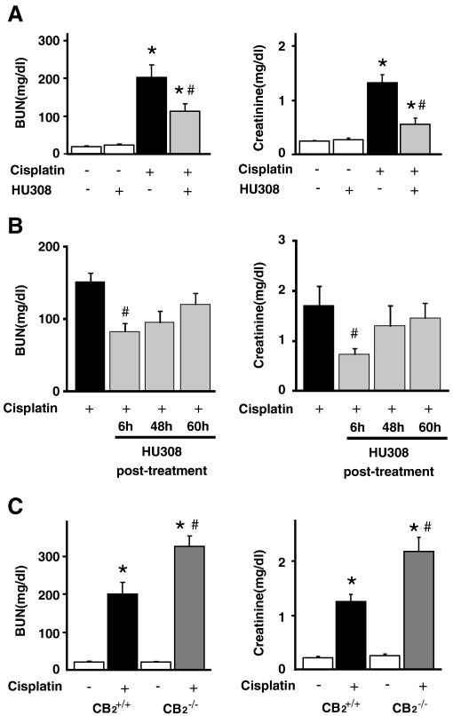
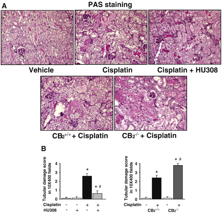
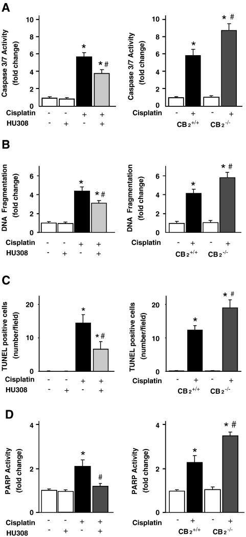
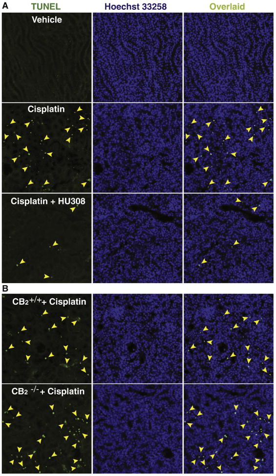
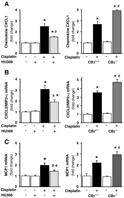
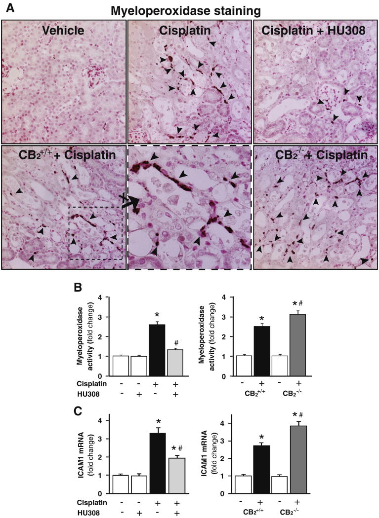
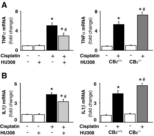
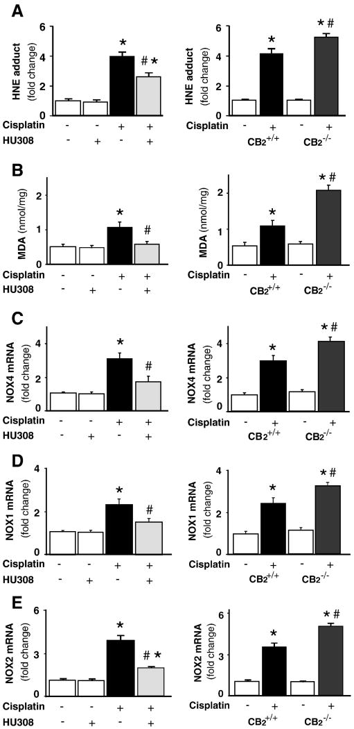
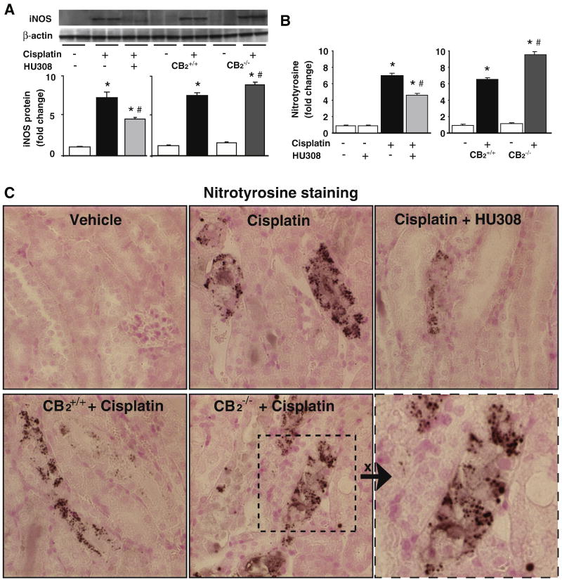
Cisplatin is an important chemotherapeutic agent; however, its nephrotoxicity limits its clinical use. Enhanced inflammatory response and oxidative/nitrosative stress seem to play a key role in the development of cisplatin-induced nephropathy. Activation of cannabinoid-2 (CB(2)) receptors with selective agonists exerts anti-inflammatory and tissue-protective effects in various disease models. We have investigated the role of CB(2) receptors in cisplatin-induced nephrotoxicity using the selective CB(2) receptor agonist HU-308 and CB(2) knockout mice. Cisplatin significantly increased inflammation (leukocyte infiltration, CXCL1/2, MCP-1, TNFalpha, and IL-1beta levels) and expression of adhesion molecule ICAM-1 and superoxide-generating enzymes NOX2, NOX4, and NOX1 and enhanced ROS generation, iNOS expression, nitrotyrosine formation, and apoptotic and poly(ADP-ribose) polymerase-dependent cell death in the kidneys of mice, associated with marked histopathological damage and impaired renal function (elevated serum BUN and creatinine levels) 3 days after the administration of the drug. CB(2) agonist attenuated the cisplatin-induced inflammatory response, oxidative/nitrosative stress, and cell death in the kidney and improved renal function, whereas CB(2) knockouts developed enhanced inflammation and tissue injury. Thus, the endocannabinoid system, through CB(2) receptors, protects against cisplatin-induced kidney damage by attenuating inflammation and oxidative/nitrosative stress, and selective CB(2) agonists may represent a promising novel approach to preventing this devastating complication of chemotherapy.


Conflict of interest statement

Disclosures: No conflict of interest to disclose.

Figures

Figure 1
Figure 1. CB2 activation attenuates the cisplatin-induced renal dysfunction; enhanced cisplatin-induced dysfunction in CB2 knockout mice
Cisplatin induced profound renal dysfunction 72 h after the administration to mice evidenced by increased serum levels of blood urea nitrogen (BUN) and creatinine (Panel A), which were attenuated by CB2 agonist HU-308 treatment (starting from 1.5 hrs before the cisplatin injection and every 24 hours thereafter until the measurements were taken at 72 hours). Starting from 6 hrs after the cisplatin administration HU-308 was still effective, however it lost efficacy when it was administered after the full inflammatory response already developed at 48 or 60 hours (panel B). Results are mean±S.E.M. of n=8-10/group, *P<0.01 vs. vehicle; #P<0.05 vs. cisplatin). The cisplatin-induced renal dysfunction was enhanced in CB2-/- mice compared to CB2+/+ littermates (Panel C). Results are mean±S.E.M. of 6-7 experiments/group *P<0.01 vs. vehicle in CB2+/+ or CB2-/- mice; #P<0.05 vs. cisplatin in CB2+/+ mice.
Figure 2
Figure 2. CB2 activation attenuates the cisplatin-induced renal histopathological damage; enhanced cisplatin-induced damage in CB2 knockout mice
As shown in the representative images (panel A) cisplatin induced profound histopathological renal injury 72 h after the administration to mice, evidenced by protein cast, vacuolation and desquamation of epithelial cells in the renal tubules using PAS staining, which were attenuated by HU-308 treatment starting from 1.5 hrs before the cisplatin injection (Panels A upper row, panel B left). The cisplatin-induced renal damage was enhanced in CB2-/- mice compared to CB2+/+ littermates (Panels A bottom row, panel B right). Results are mean±S.E.M. of 6-7 experiments/group *P<0.01 vs. vehicle in CB2+/+ or CB2-/- mice; #P<0.01 vs. cisplatin in CB2+/+ mice.
Figure 3
Figure 3. CB2 activation attenuates the cisplatin-induced renal cell death; enhanced cisplatin-induced cell death in CB2 knockout mice
Cell death in the kidneys was evaluated by caspase 3/7 activity, DNA fragmentation and PARP activity assays (Panels A-D). As shown in panels 3A-D left columns, all markers of cisplatin-induced cell death in the kidneys were attenuated by HU-308 treatment starting from 1.5 hrs before the cisplatin injection. Results are mean±S.E.M. of 5-7 experiments in each group *P<0.01 vs. vehicle; #P<0.05 vs. The cisplatin-induced cell death was enhanced in kidneys of CB2-/- mice compared to CB2+/+ littermates (Panels A-D right column). Results are mean±S.E.M. of 6-7 experiments/group *P<0.01 vs. vehicle in CB2+/+ or CB2-/- mice; #P<0.01 vs. cisplatin in CB2+/+ mice.
Figure 4
Figure 4. CB2 activation attenuates the cisplatin-induced renal TUNEL staining; enhanced cisplatin-induced TUNEL staining in kidneys of CB2 knockout mice
Late apoptotic cell death in kidneys was evaluated by fluorescent TUNEL staining (Panels A and B, left column). As shown in representative images (320×magnification), cisplatin markedly increased the number of TUNEL positive cells (left column, shown in green color) in the kidneys. Middle column depicts nuclear staining with Hoechst 33258 (blue color), and the collocalization is shown with TUNEL at right. The number of TUNEL positive cells was attenuated by HU-308 treatment starting from 1.5 hrs before the cisplatin injection (Panel A). The cisplatin-induced cell death (number of TUNEL positive cells) was increased in kidneys of CB2-/- mice compared to CB2+/+ littermates (Panel B). See also Figure 3C for quantification.
Figure 5
Figure 5. CB2 activation attenuates the cisplatin-induced enhanced renal chemokine mRNA expressions, increased cisplatin-induced chemokine expression in CB2 knockout mice
Cisplatin significantly increased chemokine (C-X-C motif) ligand 1 (CXCL1 or KC; Panel A), chemokine (C-X-C motif) ligand 2 (CXCL2 or macrophage-inflammatory protein-2 (MIP-2); Panel B), monocyte chemoattractant protein-1 (MCP-1, Panel C) mRNA expressions in the kidneys (Panels A-C), which were attenuated by CB2 agonist HU-308 treatment (starting from 1.5 hrs before the cisplatin injection; Panels 5A-C, left panels). Results are mean±S.E.M. of 5-10 experiments in each group *P<0.01 vs. vehicle; #P<0.05 vs. cisplatin. The cisplatin-induced increased chemokine expressions were enhanced in kidneys of CB2-/- mice compared to CB2+/+ littermates (Panels A-C right panels). Results are mean±S.E.M. of 6-7 experiments/group *P<0.01 vs. vehicle in CB2+/+ or CB2-/- mice; #P<0.05 vs. cisplatin in CB2+/+ mice.
Figure 6
Figure 6. CB2 activation attenuates the cisplatin-induced increased renal leukocyte infiltration and adhesion molecule ICAM-1 expression; enhanced cisplatin-induced inflammation in CB2 knockout mice
Cisplatin significantly increased the number of renal myeloperoxidase positive cells around the damaged tubular structures (Panel A; 400× magnification) and/or tissue myeloperoxidase activity (indicator of leukocyte infiltration, Panel B) and ICAM-1 expression (Panel C), which were attenuated by CB2 agonist HU-308 treatment (starting from 1.5 hrs before the cisplatin injection). Results are mean±S.E.M. of 5-10 experiments in each group *P<0.01 vs. vehicle; #P<0.05 vs. cisplatin. The cisplatin-induced increased renal leukocyte infiltration and ICAM-1 expression were enhanced in kidneys of CB2-/- mice compared to CB2+/+ littermates (Panel A lower row of images; Panels B and C, right panels). Results are mean±S.E.M. of 6-7 experiments/group *P<0.01 vs. vehicle in CB2+/+ or CB2-/- mice; #P<0.05 vs. cisplatin in CB2+/+ mice.
Figure 7
Figure 7. CB2 activation attenuates the cisplatin-induced increased expression of mRNA of TNF-α and IL1β; enhanced cisplatin-induced inflammatory response in CB2 knockout mice
Cisplatin significantly increased mRNA expression of TNF-α and IL1β (panels A and B) in the kidneys 72 h following the administration to mice, which were attenuated by HU-308 treatment (starting from 1.5 hrs before the cisplatin injection). Results are mean±S.E.M. of 5-10 experiments in each group *P<0.01 vs. vehicle; #P<0.05 vs. cisplatin. The cisplatin-induced increased renal TNF-α and IL1β mRNA expression was enhanced in kidneys of CB2-/- mice compared to CB2+/+ littermates (Panel A and B, right panels). Results are mean±S.E.M. of 6-7 experiments/group *P<0.01 vs. vehicle in CB2+/+ or CB2-/- mice; #P<0.05 vs. cisplatin in CB2+/+ mice.
Figure 8
Figure 8. CB2 activation attenuates the cisplatin-induced increased lipid peroxidation/ROS generation, overexpression of superoxide generating enzymes NOX4(RENOX), NOX2(gp91phox) and NOX1; increased cisplatin-induced ROS generation in CB2 knockout mice
Cisplatin induced marked increases in lipid peroxidation/ROS generation (HNE and MDA; panela A and B); mRNA expression of NOX4, NOX2 and NOX1 (Panels C-E) in the kidneys 72 h following the administration to mice, which were attenuated by HU-308 treatment (starting from 1.5 hrs before the cisplatin injection, Panels A and B, left). Results are mean±S.E.M. of 5-10 experiments in each group *P<0.01 vs. vehicle; #P<0.05 vs. cisplatin. The cisplatin-induced increased renal NOX1, NOX2 and NOX4 mRNA expression was enhanced in kidneys of CB2-/- mice compared to CB2+/+ littermates (Panels C-E, right). Results are mean±S.E.M. of 6-7 experiments/group *P<0.01 vs. vehicle in CB2+/+ or CB2-/- mice; #P<0.05 vs. cisplatin in CB2+/+ mice.
Figure 9
Figure 9. CB2 activation attenuates the cisplatin-induced enhanced renal expression of iNOS and 3-nitrotyrosine (NT) formation; increased cisplatin-induced iNOS expression and NT formation in CB2 knockout mice
Cisplatin increased iNOS protein expression (A) and NT formation (B) in the kidneys 72 h following the administration to mice, which were attenuated by HU-308 treatment (starting from 1.5 hrs before the cisplatin injection). Results are mean±S.E.M. of 5-7 experiments in each group *P<0.01 vs. vehicle; #P<0.05 vs. cisplatin. The cisplatin-induced increased renal iNOS expression and NT formation was enhanced in kidneys of CB2-/- mice compared to CB2+/+ littermates (Panels A and B, right). Results are mean±S.E.M. of 6-7 experiments/group *P<0.01 vs. vehicle in CB2+/+ or CB2-/- mice; #P<0.05 vs. cisplatin in CB2+/+ mice. Representative kidney sections demonstrate that the NT staining (dark staining) is predominantly localized in damaged tubular cells and to a lesser extent to inflammatory cells at close proximity. 400× magnification

References

    1. Ries F, Klastersky J. Nephrotoxicity induced by cancer chemotherapy with special emphasis on cisplatin toxicity. Am J Kidney Dis. 1986;8:368–379. - PubMed
    1. Schrier RW. Cancer therapy and renal injury. J Clin Invest. 2002;110:743–745. - PMC - PubMed
    1. Yamate J, Sato K, Ide M, Nakanishi M, Kuwamura M, Sakuma S, Nakatsuji S. Participation of different macrophage populations and myofibroblastic cells in chronically developed renal interstitial fibrosis after cisplatin-induced renal injury in rats. Vet Pathol. 2002;39:322–333. - PubMed
    1. Faubel S, Lewis EC, Reznikov L, Ljubanovic D, Hoke TS, Somerset H, Oh DJ, Lu L, Klein CL, Dinarello CA, Edelstein CL. Cisplatin-induced acute renal failure is associated with an increase in the cytokines interleukin (IL)-1beta, IL-18, IL-6, and neutrophil infiltration in the kidney. J Pharmacol Exp Ther. 2007;322:8–15. - PubMed
    1. Matsushima H, Yonemura K, Ohishi K, Hishida A. The role of oxygen free radicals in cisplatin-induced acute renal failure in rats. J Lab Clin Med. 1998;131:518–526. - PubMed

Publication types

MeSH terms