Spectrum of CD30+ lymphoid proliferations in the eyelid lymphomatoid papulosis, cutaneous anaplastic large cell lymphoma, and anaplastic large cell lymphoma
- PMID: 19969358
- PMCID: PMC2830810
- DOI: 10.1016/j.ophtha.2009.07.013
Spectrum of CD30+ lymphoid proliferations in the eyelid lymphomatoid papulosis, cutaneous anaplastic large cell lymphoma, and anaplastic large cell lymphoma
Abstract
Purpose: To report the clinicopathologic features of 3 patients with CD30(+) lymphoid proliferations of the eyelid.
Design: Retrospective case series.
Participants: Patients with cutaneous CD30(+) lymphoproliferative lesions of the eyelid.
Methods: Three patients with CD30(+) non-mycosis fungoides T-cell lymphoid infiltrates of the eyelid were identified. The histories, clinical findings, pathologic features including immunohistochemical staining, treatments, and outcomes were reviewed and compared.
Main outcome measures: Pathologic findings including immunohistochemical analysis.
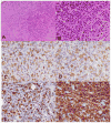
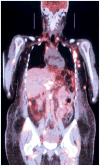
Results: The patients included an 81-year-old man, an 18-year-old man, and a 42-year-old woman with CD30(+) lymphoid proliferations of the eyelid and adjacent soft tissue. The first patient had an isolated crateriform eyelid lesion that was classified as lymphomatoid papulosis (LyP). The second patient had an isolated multinodular lesion of the eyelid that was classified as cutaneous anaplastic large cell lymphoma (cALCL). The third patient presented with eyelid edema with an underlying mass and was found to have widely disseminated anaplastic large cell lymphoma (ALCL). Diagnoses were dependent on clinical findings.
Conclusions: The CD30(+) lymphoid proliferations represent a spectrum of conditions ranging from indolent LyP, to moderately aggressive cALCL, to highly aggressive ALCL. Interpretation of the pathologic findings in CD30(+) lymphoid proliferations is based in part on clinical findings.
Financial disclosure(s): The authors have no proprietary or commercial interest in any material discussed in this article.
Copyright (c) 2010 American Academy of Ophthalmology. Published by Elsevier Inc. All rights reserved.
Figures


References
-
- Droc C, Cualing HD, Kadin ME. Need for an improved molecular/genetic classification for CD30+ lymphomas involving the skin. Cancer Control. 2007;14:124–32. - PubMed
-
- Assaf C, Hirsch B, Wagner F, et al. Differential expression of TRAF1 aids in the distinction of cutaneous CD30-positive lymphoproliferations. J Invest Dermatol. 2007;127:1898–904. - PubMed
-
- Coupland SE, Foss HD, Assaf C, et al. T-cell and T/natural killer-cell lymphomas involving ocular and ocular adnexal tissues: a clinicopathologic, immunohistochemical, and molecular study of seven cases. Ophthalmology. 1999;106:2109–20. - PubMed
-
- LeBoit PE. Lymphomatoid papulosis and cutaneous CD30+ lymphoma. Am J Dermatopathol. 1996;18:221–35. - PubMed
-
- Stein H, Mason DY, Gerdes J, et al. The expression of the Hodgkin’s disease associated antigen Ki-1 in reactive and neoplastic lymphoid tissue: evidence that Reed-Sternberg cells and histiocytic malignancies are derived from activated lymphoid cells. Blood. 1985;66:848–58. - PubMed
Publication types
MeSH terms
Substances
Grants and funding
LinkOut - more resources
Full Text Sources
Medical

