. 2010 Mar;31(3):489-95.
doi: 10.1093/carcin/bgp305. Epub 2009 Dec 7.

Dietary omega-3 polyunsaturated fatty acids suppress expression of EZH2 in breast cancer cells

Affiliations

Dietary omega-3 polyunsaturated fatty acids suppress expression of EZH2 in breast cancer cells

Manjari Dimri et al. Carcinogenesis. 2010 Mar.

Abstract

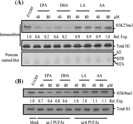
The polycomb group (PcG) protein, enhancer of zeste homologue 2 (EZH2), is overexpressed in several human malignancies including breast cancer. Aberrant expression of EZH2 has been associated with metastasis and poor prognosis in cancer patients. Despite the clear role of EZH2 in oncogenesis and therapy failure, not much is known about chemotherapeutics and chemopreventive agents that can suppress its expression and activity. Here, we show that dietary omega-3 (omega-3) polyunsaturated fatty acids (PUFAs) can regulate the expression of EZH2 in breast cancer cells. The treatment of breast cancer cells with omega-3 PUFAs, but not omega-6 PUFAs, led to downregulation of EZH2. Studies using proteosome inhibitor MG132 suggested that omega-3 PUFAs induce degradation of the PcG protein EZH2 through posttranslational mechanisms. Furthermore, downregulation of EZH2 by omega-3 PUFAs was accompanied by a decrease in histone 3 lysine 27 trimethylation (H3K27me3) activity of EZH2 and upregulation of E-cadherin and insulin-like growth factor binding protein 3, which are known targets of EZH2. Treatment with omega-3 PUFAs also led to decrease in invasion of breast cancer cells, an oncogenic phenotype that is known to be associated with EZH2. Thus, our studies suggest that the PcG protein EZH2 is an important target of omega-3 PUFAs and that downregulation of EZH2 may be involved in the mediation of anti-oncogenic and chemopreventive effects of omega-3 PUFAs.


Figures

Fig. 1.
Fig. 1.
ω-3 PUFAs but not ω-6 PUFAs downregulate EZH2 in MDA-MB-231 (A), MCF7 (B) and T47D (C) breast cancer cells. These cell lines were treated with EPA, DHA, LA or AA and analyzed for the expression of EZH2 by western blot analysis as described in Materials and Methods. The relative expression (Rel. Exp.) of EZH2 in PUFA- and mock-treated cells was determined by densitometric analysis of EZH2 signal in each lane and normalizing it to β-actin signal of the respective lane. The densitometric analysis was performed using Image J software (National Institutes of Health, Bethesda, MD).
Fig. 2.
Fig. 2.
ω-3 PUFAs posttranslationally regulate the expression of EZH2. (A) RT–PCR analysis of mock-, EPA-, DHA-, LA- and AA-treated MDA-MB-231 cells was performed using primers specific for EZH2 and β-actin as described in Materials and Methods. The relative expression of EZH2 transcript was determined by densitometry as described in Figure 1. (B) MDA-MB-231 cells were either pretreated with MG132 for 60 min or mock-treated with dimethyl sulfoxide (solvent), followed by treatment with EtOH, EPA, DHA, LA and AA for 8 h in the presence or absence of MG132. After treatment, western blot analysis for EZH2 and β-actin was performed and the relative expression of EZH2 was determined by densitometry as described in Figure 1. (C) MDA-MB-231 cells were mock treated or DHA treated in the presence or absence of MG132 for 8 h, and IP was performed using a mAb against EZH2 and immunoglobulin G (negative control). Immunoblot (IB) analysis was performed using anti-ubiquitin and anti-EZH2 antibodies. Of the total extract used for IP, 10% of each sample was also analyzed for EZH2 input. (D) Half-life of EZH2 in mock- and DHA-treated (for 8 h) MDA-MB-231 cells was determined by blocking synthesis of new proteins using 100 μg/ml cyclohexamide (CHX) at different time points (0–90 min) and rate of EZH2 proteolysis was determined by western blot analysis of remaining EZH2 protein at each time point and normalizing it to β-actin using densitometry as described in Figure 1 and Materials and Methods.
Fig. 3.
Fig. 3.
ω-3 PUFAs, but not ω-6 PUFAs, downregulate H3K27me3 and H3K9me3 in MDA-MB-231 cells. (A) Total histones were extracted from mock- or EPA-, DHA-, LA- and AA-treated cells (8 h) using acid extraction as described in Materials and Methods. Ponceau staining of the membrane was done to visualize total histones, and western blot analysis was performed to detect total H3 and H3K27me3 levels. (B) Total histones were analyzed for the expression of H3K9me3 and total H3 by western blot analysis.
Fig. 4.
Fig. 4.
ω-3 PUFAs, but not ω-6 PUFAs, downregulate H3K27me3, which correlates with EZH2 downregulation by ω-3 PUFAs in MDA-MB-231 cells. Cells were treated with 40 μM PUFAs or mock treated with EtOH in chamber slides, fixed and coimmunostained using antibodies specific for EZH2 (BD Biosciences) and H3K27me3 (Upstate Biotechnology) and stained with nuclear stain 4′,6-diamidino-2-phenylindole (DAPI). After staining, cells were photographed (×40) using a Nikon Eclipse 80i confocal microscope and colocalization was studied by merging different colors [Alexa Fluor 488 (EZH2), Alexa Fluor 594 (H3K27me3) and DAPI].
Fig. 5.
Fig. 5.
ω-3 PUFAs upregulate E-cadherin and IGFBP3 and inhibits invasion in MDA-MB-231 cells. (A) Upregulation of E-cadherin by DHA in MDA-MB-231 cells was determined by the western blot analysis using an antibody specific to E-cadherin (BD Biosciences). (B) Equal number of MDA-MB-231 cells were plated and treated with EtOH or PUFAs for 8 h in equal volume of cell culture medium. IGFBP3 expression in the trichloroacetic acid-precipitated supernatant of treated cells was determined by western blot analysis using an anti-IGFBP3 antibody (Santa Cruz Biotechnology) as described in Materials and Methods. Ponceau staining of the blot was done to confirm the loading of supernatant proteins. The major band in the Ponceau stained blot probably represents immunoglobulin G from the serum constituent of the medium. (C) Effect of ω-3 and ω-6 PUFAs on invasion in MDA-MB-231 cells was determined by Matrigel Invasion assays using 24-well BD Biocoat Matrigel Invasion Chambers (BD Biosciences) as described in Materials and Methods. Columns indicates mean of triplicate samples; bars represents SD; *P < 0.002, **P < 0.003.

References

    1. Pasini D, et al. Polycomb group proteins in cell cycle progression and cancer. Cell Cycle. 2004;3:396–400. - PubMed
    1. Sparmann A, et al. Polycomb silencers control cell fate, development and cancer. Nat. Rev. Cancer. 2006;6:846–856. - PubMed
    1. Bracken AP, et al. Genome-wide mapping of Polycomb target genes unravels their roles in cell fate transitions. Genes Dev. 2006;20:1123–1136. - PMC - PubMed
    1. Ringrose L. Polycomb comes of age: genome-wide profiling of target sites. Curr. Opin. Cell Biol. 2007;19:290–297. - PubMed
    1. Ding L, et al. Identification of EZH2 as a molecular marker for a precancerous state in morphologically normal breast tissues. Cancer Res. 2006;66:4095–4099. - PubMed

Publication types

MeSH terms