. 1991 Feb 22;64(4):789-800.
doi: 10.1016/0092-8674(91)90508-v.

Mutations in the CDP-choline pathway for phospholipid biosynthesis bypass the requirement for an essential phospholipid transfer protein

Affiliations

Mutations in the CDP-choline pathway for phospholipid biosynthesis bypass the requirement for an essential phospholipid transfer protein

A E Cleves et al. Cell. .

Abstract

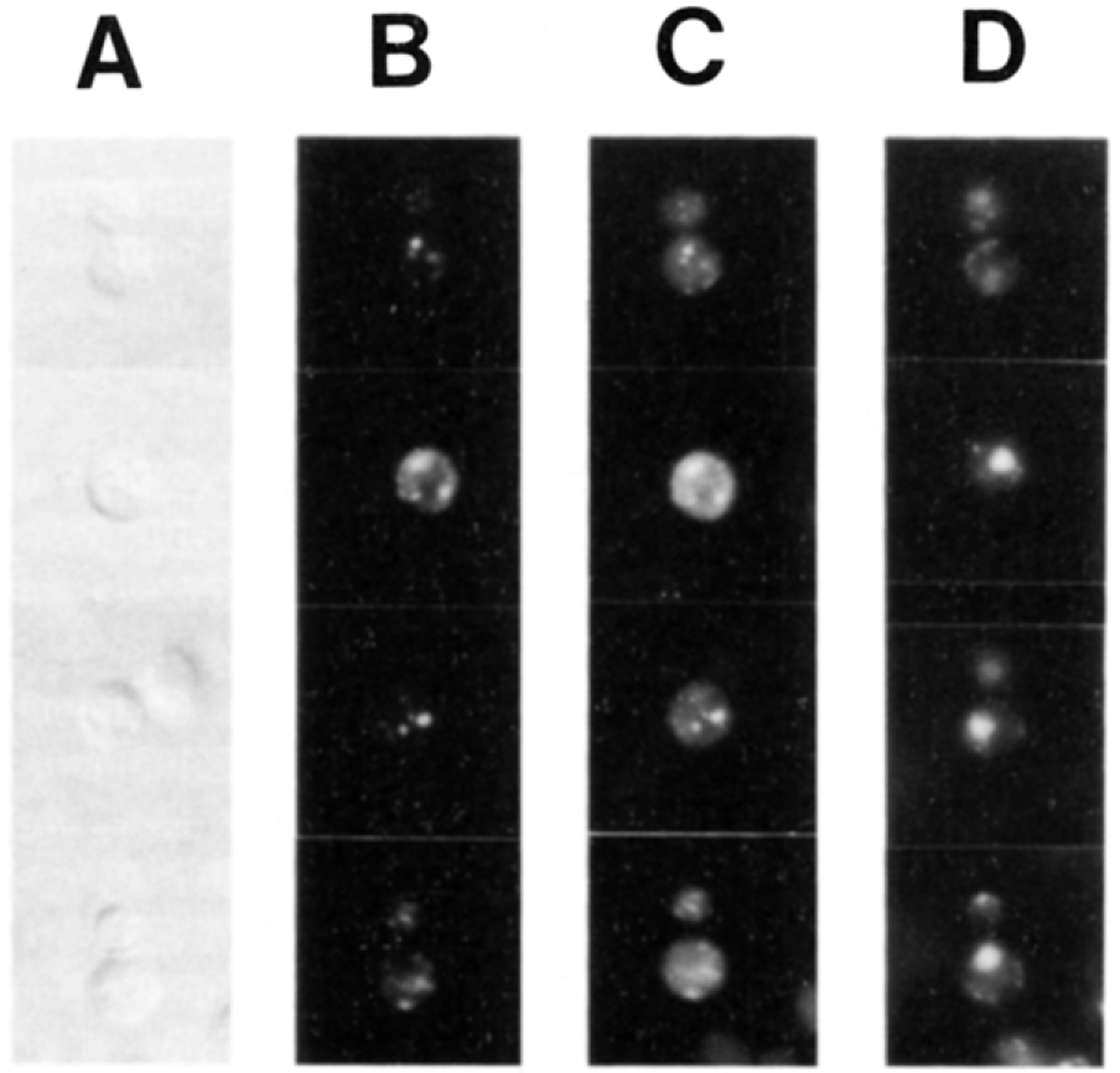
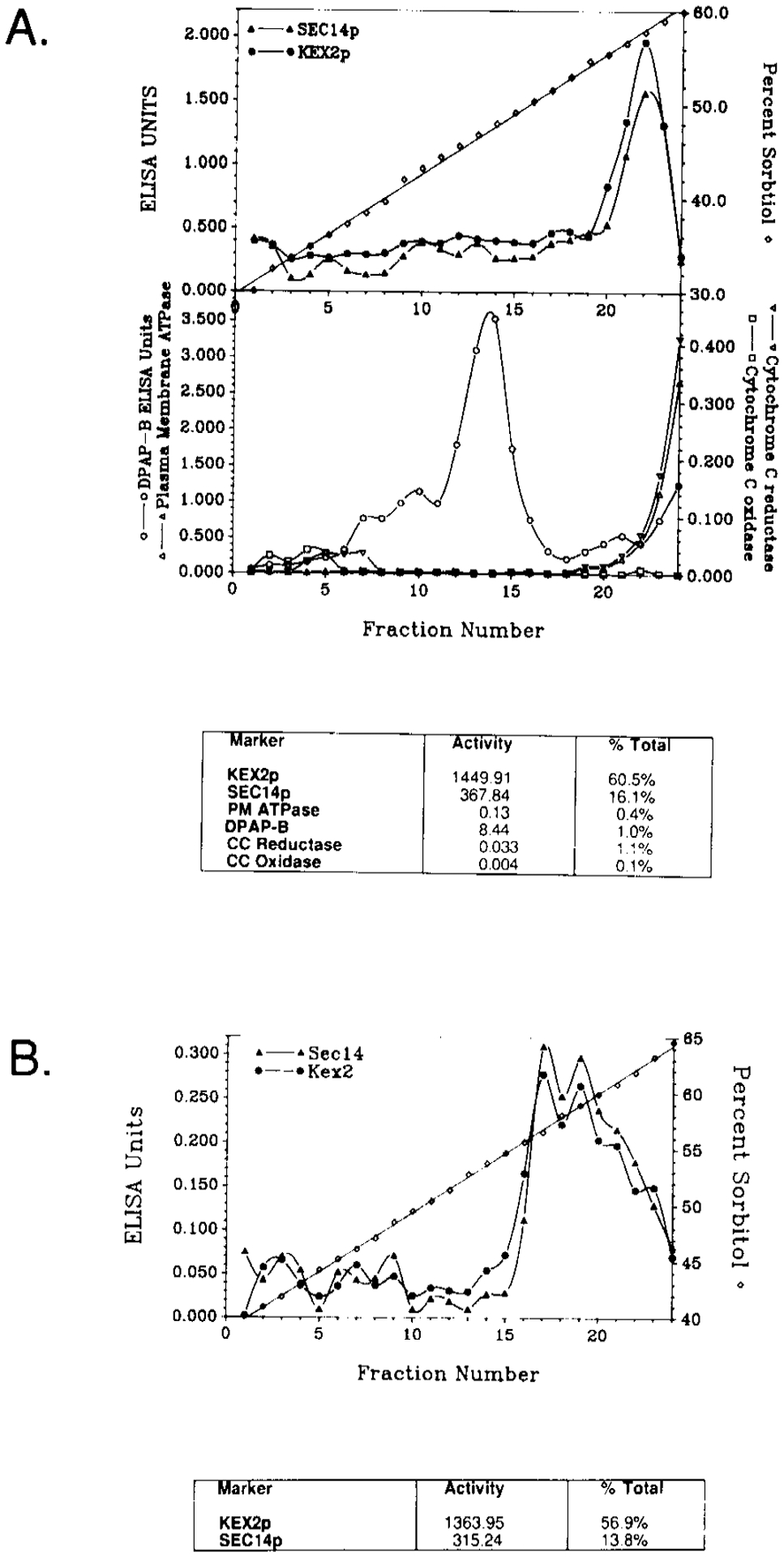
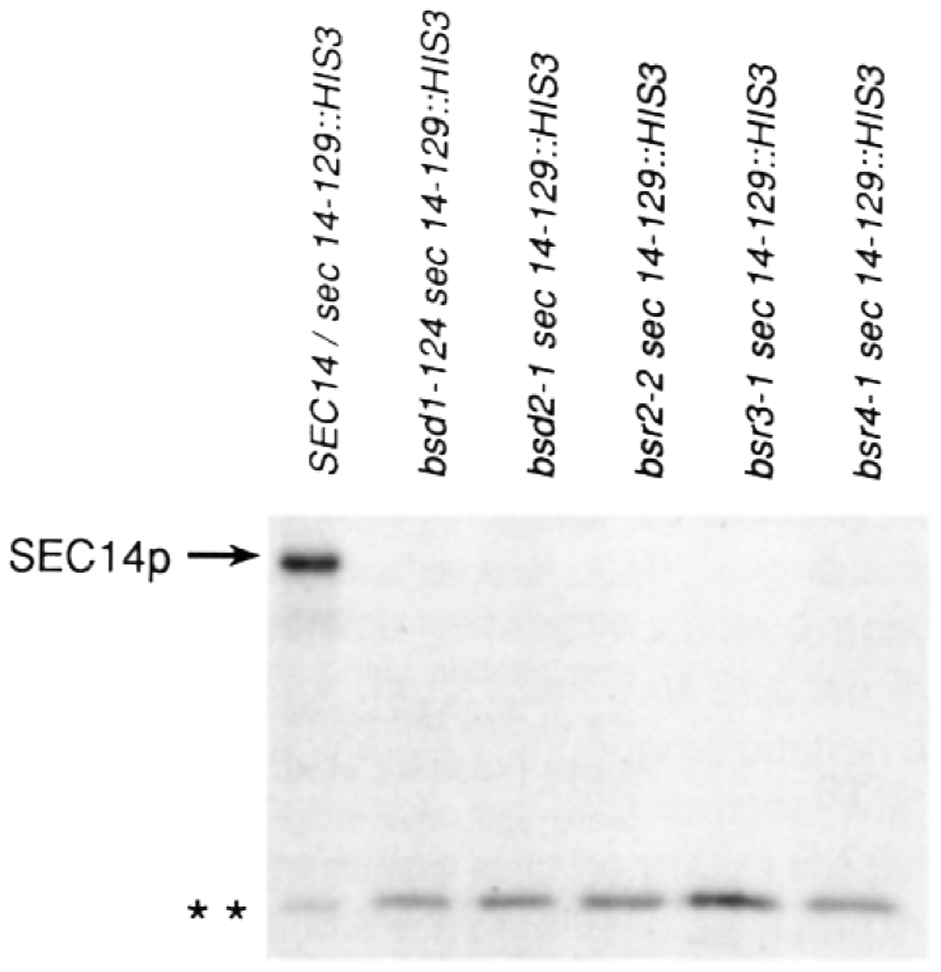
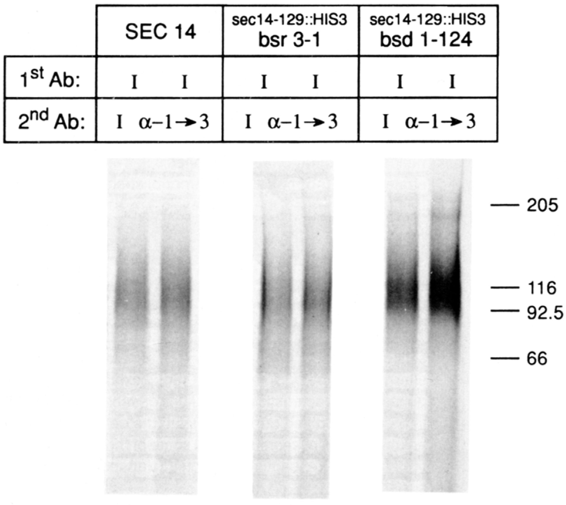
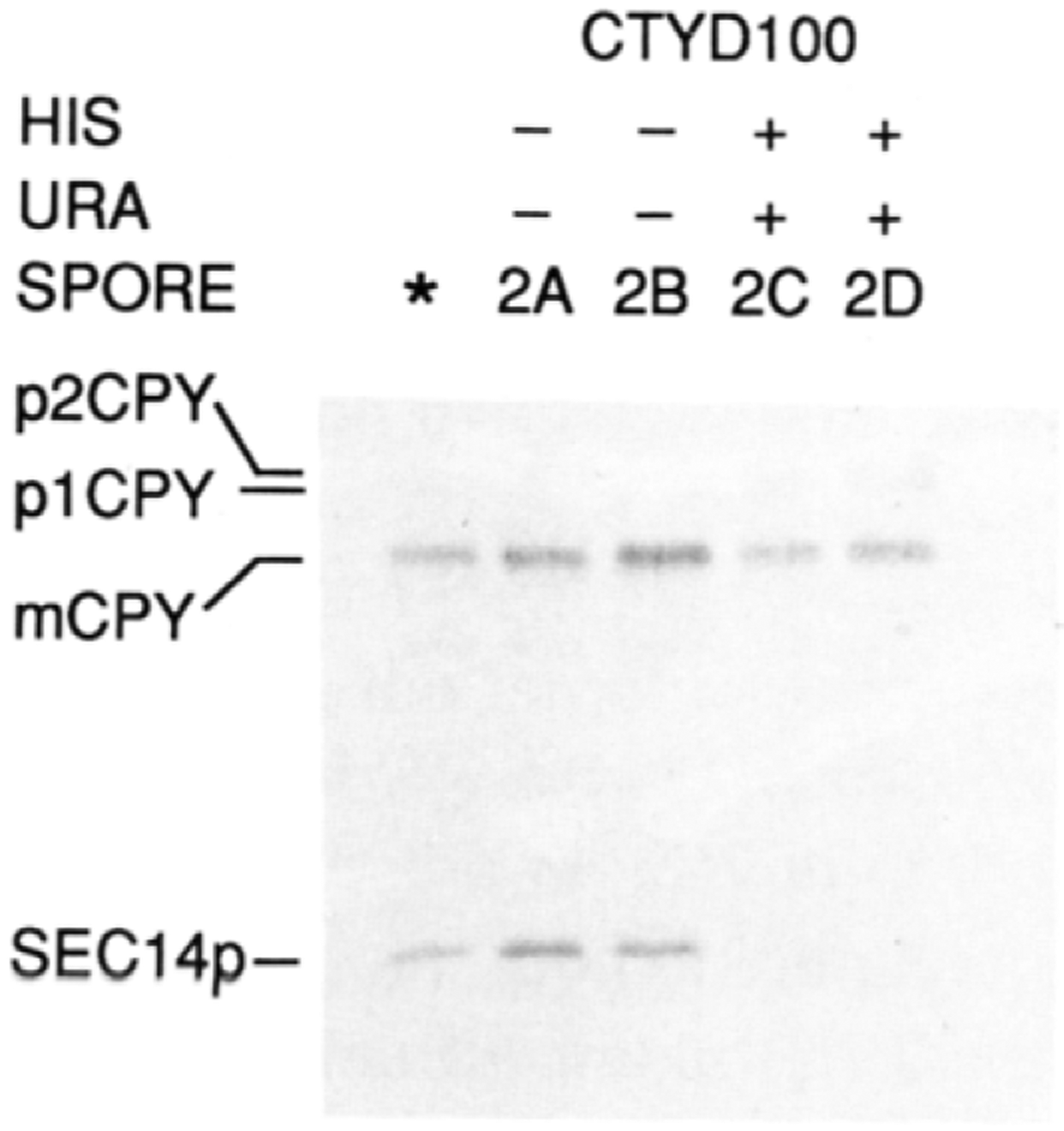
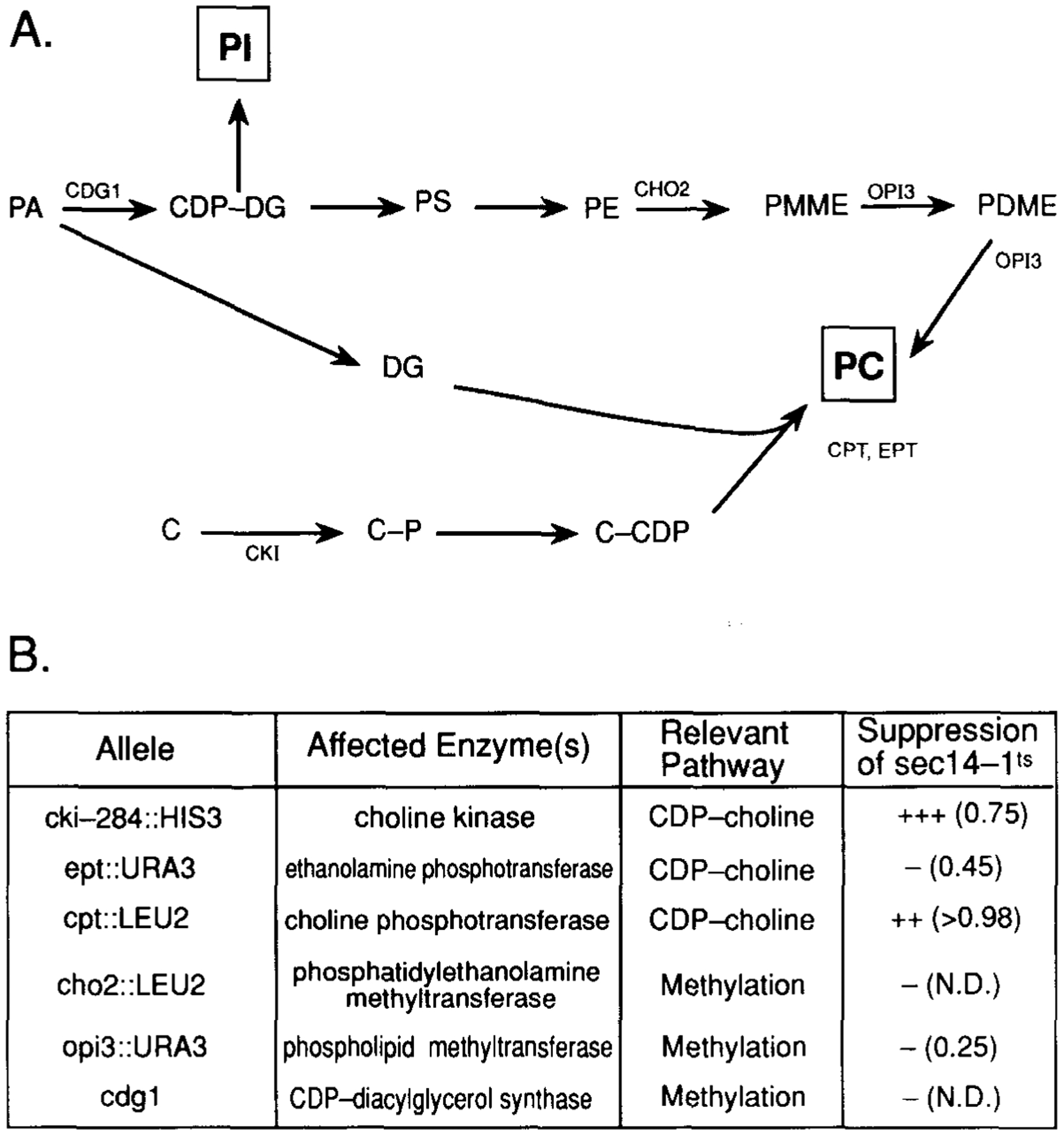
SEC14p is the yeast phosphatidylinositol (PI)/phosphatidylcholine (PC) transfer protein, and it effects an essential stimulation of yeast Golgi secretory function. We now report that the SEC14p localizes to the yeast Golgi and that the SEC14p requirement can be specifically and efficiently bypassed by mutations in any one of at least six genes. One of these suppressor genes was the structural gene for yeast choline kinase (CKI), disruption of which rendered the cell independent of the normally essential SEC14p requirement. The antagonistic action of the CKI gene product on SEC14p function revealed a previously unsuspected influence of biosynthetic activities of the CDP-choline pathway for PC biosynthesis on yeast Golgi function and indicated that SEC14p controls the phospholipid content of yeast Golgi membranes in vivo.


Figures

Figure 1.
Figure 1.
SECl4p Colocalizes with the Yeast Golgi Complex In Vivo Wild-type yeast cells were grown at 25°C and double-stained for fluorescence microscopy with TRITC-conjugated antibodies directed against antibodies bound to KEX2p, a Golgi marker, and FITC-conjugated antibodies directed against SEC14p. Cell outlines are revealed by Nomarski (A). The KEX2p (B) and SEC14p (C) staining profiles are also shown. Nuclear fluorescence is shown in (D) as determined by DAPI (4′, 6′-diamidino-2-phenylindole) staining.
Figure 2.
Figure 2.. Distribution of Marker Proteins across Gradient Fractions
(A) The P100 fraction was collected and loaded under a 30%–60% self-forming sorbitol flotation gradient. After centrifugation to equilibrium, 0.5 ml fractions were collected and assayed for the indicated markers as described in the legend to Table 1 and the Experimental Procedures. Unit values are expressed on a per fraction basis. The relative quantities of each marker in the peak KEX2p fraction pool are tabulated both in terms of units per fraction and percent of total. (B) The KEX2p fraction pool was layered upon a 40%–65% sorbitol sedimentation gradient. The gradient was fractionated and assayed for KEX2p and SEC14p by quantitative ELISA. Again, the unit values for KEX2p and SEC14p are tabulated in terms of units per fraction and percent of total.
Figure 3.
Figure 3.. Suppression of a sec14 Disruption Allele
The appropriate yeast strains were grown to early logarithmic growth phase (OD600 = 0.5) in glucose (2%) minimal medium at 30°C. Yeast cells (0.5 OD600 units) were radiolabeled for 20 min with 100 μCi of Trans-Label (>1000 Ci/mmol; ICN Radiochemicals, Irvine, CA), and radiolabeling was terminated by addition of trichloroacetic acid (5% final). SEC14p immunoreactive species were specifically precipitated and resolved by SDS-PAGE and autoradiography as previously described (Bankaitis et al., 1989; Salama et al., 1990). The relevant genotypes of the corresponding yeast strains are indicated above (strains CTYD43, CTY221, CTY217, CTY228, CTY229, and CTY230, respectively). Complete genotypes for these strains are given in Table 4. The positions of the full-length SEC14p and the truncated sec14129::HIS3 (* *) gene product are indicated at left. The immunoprecipitates were normalized on the basis of total incorporated cpm in the clarified extracts. The samples obtained from the suppressor strains were severalfold overloaded with respect to those obtained from the diploid strains.
Figure 4.
Figure 4.. Invertase Glycosylation in the Suppressor Strains
The appropriate yeast strains were grown to early logarithmic growth phase (OD600 = 0.5) in glucose (2%) minimal medium, washed two times with distilled water, and resuspended in glucose (0.1%) minimal medium. After a 2 hr incubation at 25°C to allow derepression of invertase synthesis, the cells (2 OD600 units) were shifted to 37°C and allowed to incorporate radiolabel (250 μCi of Trans-Label) for 1 hr. Invertase antigen was precipitated from clarified extracts by an initial round (1stAb) of immunoprecipitation using invertase-specific antibodies (I) directed solely against polypeptide epitopes (Klionsky et al., 1988). The precipitates were split and subjected to a second round (2ndAb) of immunoprecipitation with either the control invertase sera (I) or the test antibodies (α-1→3) raised specifically against α-1→3 mannose linkages of outer chain carbohydrate (Franzusoff and Schekman, 1989). The relevant genotypes of the yeast strains employed (i.e., CTY182, CTY229, and CTY221, respectively) are given at the top of the corresponding lanes, as are the designations for the 2nd Ab used. Precipitates were resolved on SDS gels that were 7.5% in acrylamide.
Figure 5.
Figure 5.. Suppression of sec14–88::URA3 by a cki Disruption Allele
The radiolabeled CPY and SEC14p profiles from meiotic progeny derived from an ascus that exhibited four viable spores, in a cross where sec14–88::URA3 and cki-284::HIS3 were segregating, are shown. Also included are the profiles obtained for the parental diploid strain CTYD100 (*). The details of the cross are given in the text. The meiotic progeny were cultured in glucose (2%) minimal medium, to an OD600 of approximately 0.5, at 37°C. A 30 min labeling period (100 μCi of Trans-Label) followed and was terminated with trichloroacetic acid. SEC14p and CPY immunoreactive materials were recovered and visualized after SDS-PAGE and autoradiography. The positions of the SEC14p, CPY, and p1 and p2 proCPY forms are indicated at the left. The Ura and His phenotypes of the meiotic progeny are indicated above the corresponding lanes. Further details are provided in the text.
Figure 6.
Figure 6.. Suppression of sec14 Defects by Mutations in the CDP-Choline Pathway for PC Biosynthesis
(A) The two pathways for PC biosynthesis in yeast are diagrammed, and the gene designation for structural enzymes of each pathway are indicated at the appropriate site(s) of action (for a review see Carman and Henry, 1989). Abbreviations: PA, phosphatidic acid; CDP-DG, CDP-diacylglycerol; DG, diacylglycerol; PI, phosphatidylinositol; PS, phosphatidylserine; PE, phosphatidylethanolamine; PMME, phosphatidylmonomethylethanolamine; PDME, phosphatidyldimethylethanolamine; PC, phosphatidylcholine; C, choline; C-P, choline-phosphate; C-CDP, CDP-choline. (B) The mutations in PC biosynthesis that were tested for suppression of sec14–1ts are listed. Also given are the structural enzyme and PC biosynthetic pathway affected by each mutation. Suppression was determined by constructing sec14–1ts haploid yeast strains carrying each designated mutant allele. The relevant crosses involved: meiotic analysis of CTYD100, HJ051 × CTY76, CTY214 transformed with cpt1::LEU2 as described (Hjelmstad and Bell, 1987), CTY215 × CTY410, CTY68 × CTY411, and CTY2–1C × CTY416, respectively. Complete genotypes of these strains are presented in Table 4. Phenotypic suppression was followed by testing for growth and formation of single colonies on YPD agar at 37°C, and is qualitatively indicated. The secretion index, where determined, is given in parentheses and was calculated from the ratio of extracellular invertase:total invertase as described in the legend to Table 3. These values are to be compared with those measured in these experiments for wild-type yeast (>0.98) and sec14-1ts yeast (0.20) incubated at 37°C (data not shown). The experimental conditions were exactly as those described in the legend to Table 3. N.D. = not determined.

Similar articles

Cited by

References

    1. Aitken JF, van Heusden GPH, Temkin M, and Dowhan W (1990). The gene encoding the phosphatidylinositol transfer protein is essential for cell growth. J. Biol. Chem 265, 4711–4717. - PubMed
    1. Bankaitis VA, Malehorn DE, Emr SD, and Greene R (1989). The Saccharomyces cerevisiae SEC14 gene encodes a cytosolic factor that is required for transport of secretory proteins from the yeast Golgi complex. J. Cell Biol 108, 1271–1281. - PMC - PubMed
    1. Bankaitis VA, Aitken JF, Cleves AE, and Dowhan W (1990). An essential role for a phospholipid transfer protein in yeast Golgi function. Nature 347, 561–562. - PubMed
    1. Botstein D, Falco SC, Stewart S, Brennan M, Scherer S, Stinch-comb DI, Struhl K, and Davis R (1979). Sterile host yeast (SHY): a eukaryotic system of biological containment for recombinant DNA experiments. Gene 8, 17–24. - PubMed
    1. Bowman B, and Slayman CW (1979). The effects of vanadate on the plasma membrane ATPase of Neurospora crassa. J. Biol. Chem 254, 2928–2934. - PubMed

Publication types

MeSH terms