. 2010 Jan 15;201(2):255-63.
doi: 10.1086/649589.

Toll-like receptor 2 ligand-induced protection against bacterial endophthalmitis

Affiliations

Toll-like receptor 2 ligand-induced protection against bacterial endophthalmitis

Ashok Kumar et al. J Infect Dis. .

Abstract

Background: Activation of innate immunity plays a key role in determining the outcome of an infection. Here, we investigated whether Toll-like receptors (TLRs) are involved in retinal innate response and explored the prophylactic use of TLR2 ligand in preventing bacterial endophthalmitis.

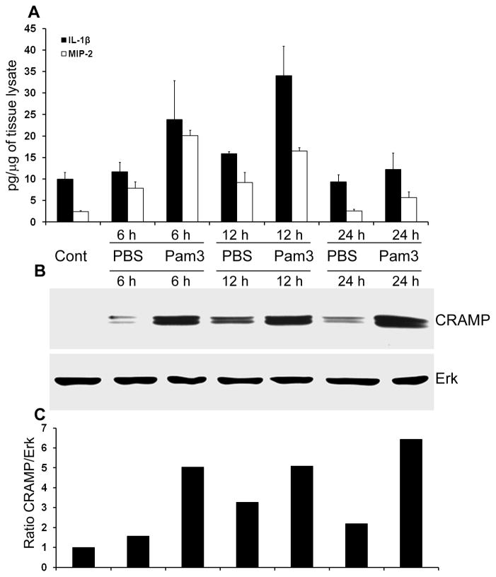
Methods: C57BL/6 mice were given intravitreal injections of Pam3Cys, a synthetic ligand of TLR2, or vehicle (phosphate-buffered saline) 24 h prior to Staphylococcus aureus inoculation. The severity of endophthalmitis was graded by slit lamp, electroretinography, histological examinations, and determination of bacterial load in the retina. The expression of cytokines/chemokines and cathelicidin-related antimicrobial peptide was assessed by enzyme-linked immunosorbent assay and Western blot, respectively.

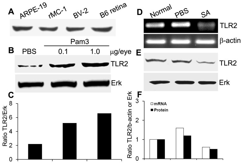
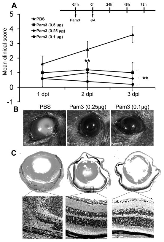
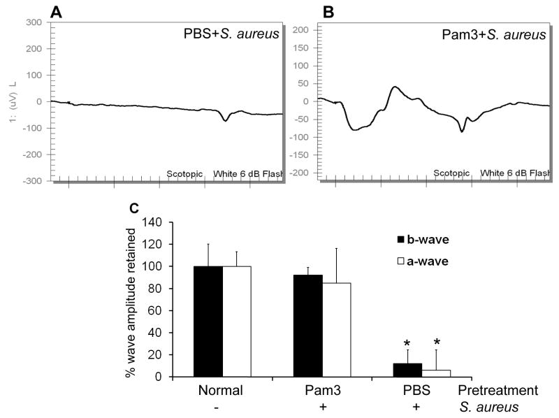
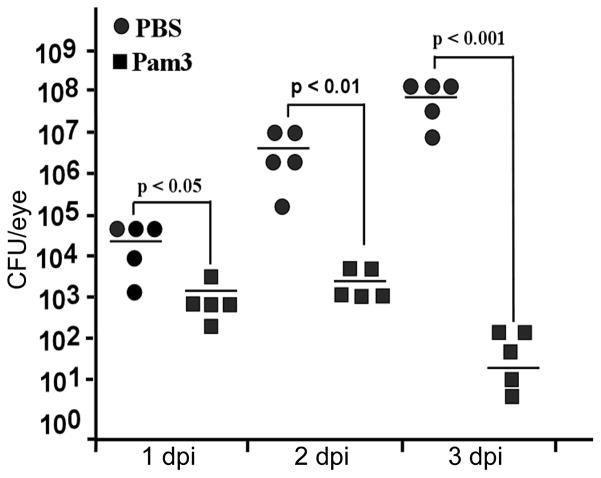
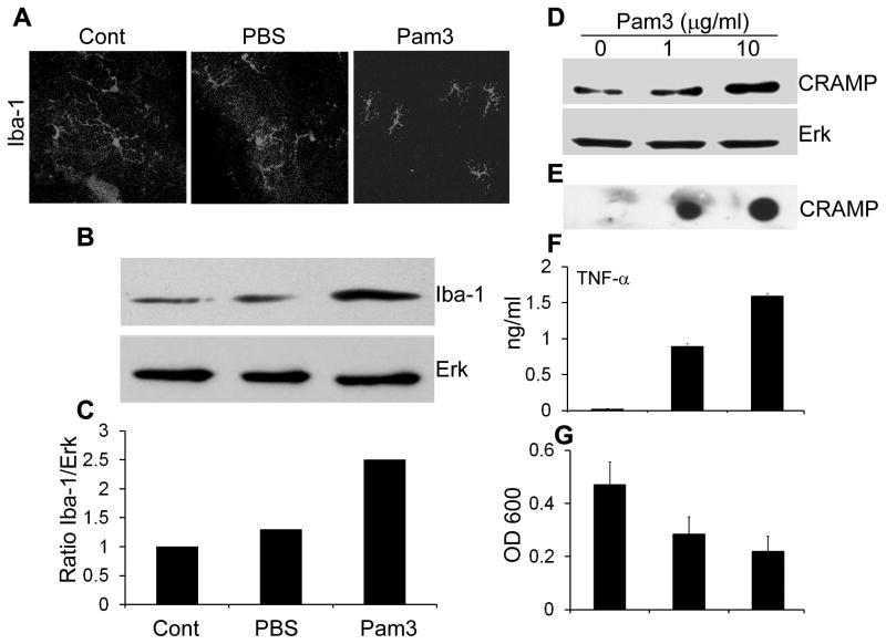
Results: Intravitreal injections of Pam3Cys up-regulated TLR2 expression in the retina of C57BL/6 mice, and Pam3Cys pretreatment significantly improved the outcome of S. aureus endophthalmitis, preserved retinal structural integrity, and maintained visual function as assessed by electroretinography in C57BL/6 mice. Furthermore, Pam3Cys pretreatment activated retinal microglia cells, induced the expression of cathelicidin-related antimicrobial peptide, and remarkably reduced the bacterial load.

Conclusions: This is the first report that highlights the existence and role of TLR2 in retinal innate immune response to S. aureus infection and suggests that modulation of TLR activation provides a novel prophylactic approach to prevent bacterial endophthalmitis.


Conflict of interest statement

Conflict of interest: Authors have no conflict of interest.

Figures

Figure 1
Figure 1. TLR2 expression in Pam3Cys and SA challenged mouse retina and retinal cells
(A) The indicated cultured cells and normal B6 mouse retina were lysed for Western blot analysis using anti-TLR2 antibody. To assess the modulation of TLR2 expression by Pam3Cys treatment in vivo, 24h after intravitreal injection of Pam3Cys (0.1 μg and 1 μg), PBS (B), and SA (E) TLR2 protein expression in whole B6 mouse retina was detected using Western blot. Expression of TLR2 mRNA at 24h post SA infection was assessed by RT-PCR in whole retina (D). The band intensity of western blot and PCR product was quantitated by densitometric analysis and normalized with their respective internal controls i.e. ERK and β-actin (C & F).
Figure 2
Figure 2. Intravitreal injection of Pam3cys prevents the development of endophthalmitis and preserves retinal architecture
(A) C57BL/6 mice (n=5 each group) were administered PBS or indicated concentrations (μg/eye) of Pam3Cys by intravitreal injection (see Methods for detail). After 24 h, eyes were inoculated with 5000 cfu of SA (RN6390). A clinical score (range 0 to 4) of Pam3Cys-pretreated and PBS injected mice on 1, 2 and 3 days post infection (dpi) was assigned for each infected eye by an ophthalmologist and presented as mean clinical scores. ANOVA was performed to compare all groups at 2 (p<0.001) and 3 dpi (p<0.001) and a nonparametric Mann-Whitney U test was performed to compare each Pam3Cys treatment to the PBS group (** p<0.001). Eyes pretreated with indicated concentration of Pam3Cys or PBS control were enucleated at 3 dpi. Light microscopy examination (B) and histopathological analysis (C) was performed on representative eyes. H&E-stained retinal tissue of PBS-pretreated and SA -infected mice showed massive infiltration as well as destruction of retinal architecture (left panel); histology of Pam3Cys-pretreated and SA-infected mice showed intact retinal architecture (right panels). Magnification, whole eyes (2.5 ×) and the retina (20 ×).
Figure 3
Figure 3. Pam3Cys pretreatment maintained normal retinal function in B6 mice eyes following SA infection
ERG response to a 6-dB flash were recorded at 3 dpi in mice were pretreated via intravitreal injection of PBS (A) or 0.1 μg of Pam3Cys (B) followed by SA infection. The percentage amplitude of a- and b-wave retained in mice pretreated with PBS or Pam3Cys were compared to normal and presented as mean ± SD ERG amplitudes (C). *P<0.001 (student’s t-test), significant decline in amplitude of a-and b-wave compared to both Pam3Cys pretreated and normal eyes.
Figure 4
Figure 4. Pam3cys pretreatment reduces bacterial load in infected B6 mice eyes
Mice were treated with PBS (n = 5) or 0.1 μg of Pam3cys/eye (n = 5) as described in the text, followed by intravitreal inoculation of 5000 cfu of SA per eye. At the indicated days of post-infection, the whole eyes were enucleated under sterile conditions, homogenized in 1ml PBS and the bacterial load was determined by serial dilution plate count. ANOVA was performed for all group at 3 dpi (p< 0.01), indicated p values were generated using paired Student’s t test.
Figure 5
Figure 5. Pam3Cys induces the expression of proinflammatory cytokines and CRAMP in the mouse retina
B6 mice were given intravitreal injection of sterile PBS or Pam3cys (0.1μg/eye). At indicated time points, the eyes (n=3 each group) were enucleated and the retinal protein lysate was used for MIP-2 and IL-1β detection by ELISA (A) and western blot analysis of CRAMP expression (B). The band intensity of western blot was quantitated by densitometric analysis and normalized with ERK, used as a protein loading control (C).
Figure 6
Figure 6. Pam3Cys induces activation of retinal microglia in vivo and production of CRAMP in vitro
(A) Mice were given intravitreal injection of sterile PBS or Pam3Cys (0.1 μg). After 24 h, the eyes were enucleated, and retinal wholemounts were prepared and stained for microglia specific marker Iba-1. Protein lysate was used for the Western blot detection of Iba-1(B) and densitometric analysis was performed after normalizing with total ERK levels (C). Cultured mouse microglia (BV-2) cells were challenged with indicated concentration of Pam3Cys. After 24 h, cell lysate was used to detect expression of CRAMP by Western blot (D), culture media was used to assess the secretion CRAMP by Dot-blot (E) and TNF-α by ELISA (F). Antibacterial activity of Pam3Cys -challenged media was assessed against SA and presented as mean ± SD of OD 600 at 8h (G).

Similar articles

Cited by

References

    1. Cebulla CM, Flynn HW., Jr Endophthalmitis after open globe injuries. Am J Ophthalmol. 2009;147:567–8. - PubMed
    1. Chiquet C, Cornut P-L, Benito Y, et al. Eubacterial PCR for Bacterial Detection and Identification in 100 Acute Postcataract Surgery Endophthalmitis. Invest Ophthalmol Vis Sci. 2008;49:1971–1978. - PubMed
    1. Krause L, Bechrakis NE, Heimann H, Kildal D, Foerster MH. Incidence and outcome of endophthalmitis over a 13-year period. Can J Ophthalmol. 2009;44:88–94. - PubMed
    1. Diago T, McCannel CA, Bakri SJ, Pulido JS, Edwards AO, Pach JM. Infectious endophthalmitis after intravitreal injection of antiangiogenic agents. Retina. 2009;29:601–5. - PubMed
    1. Scott IU, Flynn HW., Jr Reducing the risk of endophthalmitis following intravitreal injections. Retina. 2007;27:10–2. - PubMed

Publication types

Substances