

Comprehensive characterization of the DNA amplification at 13q34 in human breast cancer reveals TFDP1 and CUL4A as likely candidate target genes
- PMID: 19995430
- PMCID: PMC2815550
- DOI: 10.1186/bcr2456
Comprehensive characterization of the DNA amplification at 13q34 in human breast cancer reveals TFDP1 and CUL4A as likely candidate target genes
Abstract
Introduction: Breast cancer subtypes exhibit different genomic aberration patterns with a tendency for high-level amplifications in distinct chromosomal regions. These genomic aberrations may drive carcinogenesis through the upregulation of proto-oncogenes. We have characterized DNA amplification at the human chromosomal region 13q34 in breast cancer.
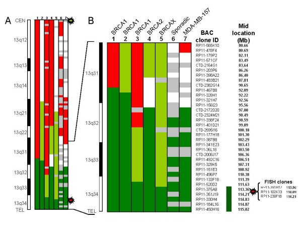
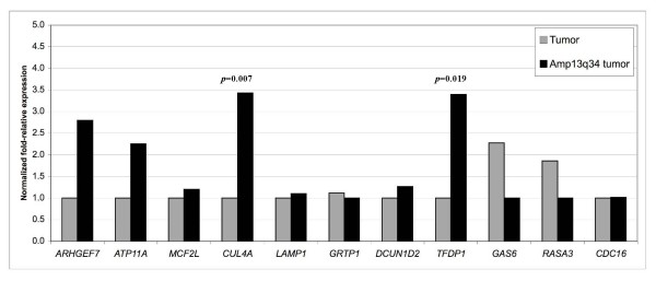
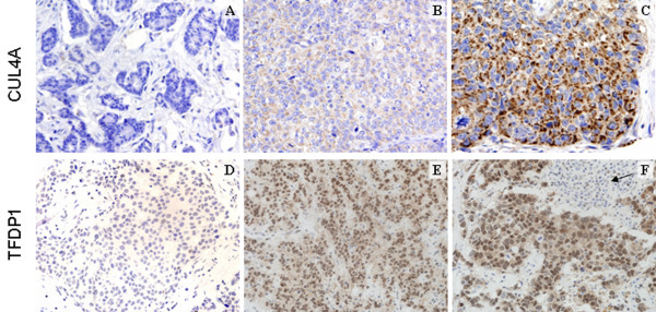
Methods: A set of 414 familial and sporadic breast cancer cases was studied for amplification at region 13q34 by fluorescence in situ hybridization (FISH) analysis on tissue microarrays. Defining the minimal common region of amplification in those cases with amplification at 13q34 was carried out using an array-based comparative genomic hybridization platform. We performed a quantitative real-time - polymerase chain reaction (qRT-PCR) gene expression analysis of 11 candidate genes located within the minimal common region of amplification. Protein expression levels of two of these genes (TFDP1 and CUL4A) were assessed by immunohistochemical assays on the same tissue microarrays used for FISH studies, and correlated with the expression of a panel of 33 antibodies previously analyzed.
Results: We have found 13q34 amplification in 4.5% of breast cancer samples, but the frequency increased to 8.1% in BRCA1-associated tumors and to 20% in basal-like tumors. Tumors with 13q34 amplification were associated with high grade, estrogen receptor negativity, and expression of EGFR, CCNE, CK5, and P-Cadherin, among other basal cell markers. We have defined a 1.83 megabases minimal common region of genomic amplification and carried out mRNA expression analyses of candidate genes located therein, identifying CUL4A and TFDP1 as the most likely target genes. Moreover, we have confirmed that tumors with 13q34 amplification significantly overexpress CUL4A and TFDP1 proteins. Tumors overexpressing either CUL4A or TFDP1 were associated with tumor proliferation and cell cycle progression markers.
Conclusions: We conclude that 13q34 amplification may be of relevance in tumor progression of basal-like breast cancers by inducing overexpression of CUL4A and TFDP1, which are both important in cell cycle regulation. Alternatively, as these genes were also overexpressed in non-basal-like tumor samples, they could play a wider role in cancer development by inducing tumor proliferation.
Figures

Similar articles
-
The CUL4A ubiquitin ligase is a potential therapeutic target in skin cancer and other malignancies.Chin J Cancer. 2013 Sep;32(9):478-82. doi: 10.5732/cjc.012.10279. Epub 2013 Jul 12. Chin J Cancer. 2013. PMID: 23845142 Free PMC article. Review.
-
Recurrent Amplification at 13q34 Targets at CUL4A, IRS2, and TFDP1 As an Independent Adverse Prognosticator in Intrahepatic Cholangiocarcinoma.PLoS One. 2015 Dec 18;10(12):e0145388. doi: 10.1371/journal.pone.0145388. eCollection 2015. PLoS One. 2015. PMID: 26684807 Free PMC article.
-
TFDP1, CUL4A, and CDC16 identified as targets for amplification at 13q34 in hepatocellular carcinomas.Hepatology. 2002 Jun;35(6):1476-84. doi: 10.1053/jhep.2002.33683. Hepatology. 2002. PMID: 12029633
-
Identification of novel amplification gene targets in mouse and human breast cancer at a syntenic cluster mapping to mouse ch8A1 and human ch13q34.Cancer Res. 2007 May 1;67(9):4104-12. doi: 10.1158/0008-5472.CAN-06-4672. Cancer Res. 2007. PMID: 17483321 Free PMC article.
-
Chromogenic and fluorescent in situ hybridization in breast cancer.Hum Pathol. 2007 Aug;38(8):1105-22. doi: 10.1016/j.humpath.2007.04.011. Hum Pathol. 2007. PMID: 17640550 Review.
Cited by
-
The CUL4A ubiquitin ligase is a potential therapeutic target in skin cancer and other malignancies.Chin J Cancer. 2013 Sep;32(9):478-82. doi: 10.5732/cjc.012.10279. Epub 2013 Jul 12. Chin J Cancer. 2013. PMID: 23845142 Free PMC article. Review.
-
Integrated analysis of genome-wide copy number alterations and gene expression in microsatellite stable, CpG island methylator phenotype-negative colon cancer.Genes Chromosomes Cancer. 2013 May;52(5):450-66. doi: 10.1002/gcc.22043. Epub 2013 Jan 23. Genes Chromosomes Cancer. 2013. PMID: 23341073 Free PMC article.
-
Dystonia, facial dysmorphism, intellectual disability and breast cancer associated with a chromosome 13q34 duplication and overexpression of TFDP1: case report.BMC Med Genet. 2013 Jul 13;14:70. doi: 10.1186/1471-2350-14-70. BMC Med Genet. 2013. PMID: 23849371 Free PMC article.
-
Targeting the protein ubiquitination machinery in melanoma by the NEDD8-activating enzyme inhibitor pevonedistat (MLN4924).Invest New Drugs. 2017 Feb;35(1):11-25. doi: 10.1007/s10637-016-0398-8. Epub 2016 Oct 25. Invest New Drugs. 2017. PMID: 27783255 Free PMC article.
-
Recurrent Amplification at 13q34 Targets at CUL4A, IRS2, and TFDP1 As an Independent Adverse Prognosticator in Intrahepatic Cholangiocarcinoma.PLoS One. 2015 Dec 18;10(12):e0145388. doi: 10.1371/journal.pone.0145388. eCollection 2015. PLoS One. 2015. PMID: 26684807 Free PMC article.
References
-
- Hu Z, Fan C, Oh DS, Marron JS, He X, Qaqish BF, Livasy C, Carey LA, Reynolds E, Dressler L, Nobel A, Parker J, Ewend MG, Sawyer LR, Wu J, Liu Y, Nanda R, Tretiakova M, Ruiz Orrico A, Dreher D, Palazzo JP, Perreard L, Nelson E, Mone M, Hansen H, Mullins M, Quackenbush JF, Ellis MJ, Olopade OI, Bernard PS, Perou CM. The molecular portraits of breast tumors are conserved across microarray platforms. BMC Genomics. 2006;7:96. doi: 10.1186/1471-2164-7-96. - DOI - PMC - PubMed
-
- Perou CM, Sorlie T, Eisen MB, Rijn M van de, Jeffrey SS, Rees CA, Pollack JR, Ross DT, Johnsen H, Akslen LA, Fluge O, Pergamenschikov A, Williams C, Zhu SX, Lonning PE, Borresen-Dale AL, Brown PO, Botstein D. Molecular portraits of human breast tumours. Nature. 2000;406:747–752. doi: 10.1038/35021093. - DOI - PubMed
-
- Bergamaschi A, Kim YH, Wang P, Sorlie T, Hernandez-Boussard T, Lonning PE, Tibshirani R, Borresen-Dale AL, Pollack JR. Distinct patterns of DNA copy number alteration are associated with different clinicopathological features and gene-expression subtypes of breast cancer. Genes Chromosomes Cancer. 2006;45:1033–1040. doi: 10.1002/gcc.20366. - DOI - PubMed
Publication types
MeSH terms
Substances
LinkOut - more resources
Full Text Sources
Other Literature Sources
Medical
Research Materials
Miscellaneous
